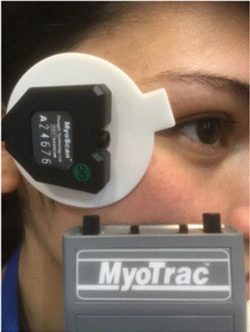
Figure 1: Portable electromyography: a useful tool to demonstrate reduction of muscular activity.

Jorge Schwember1* Luisa Madrid1 Adriana Hernández1 Fernando Leiva1 Ariel Kurtzig2
1Centro Laser, Viña del Mar, Chile*Corresponding author: Jorge Schwember, Centro Laser, Viña del Mar, Chile, E-mail: horaciosch@gmail.com
The skin is exposed to many factors that cause wrinkling and loss of elasticity. For the past 30 years, Botulinum Toxin (BT) injections have been the gold standard of treatment for wrinkle reduction. In this article, the authors propose a permanent treatment for crow’s feet wrinkles: Percutaneous Electromyoneurolysis (PEMN) of the Orbicularis Oculi Muscle (OOM) which blocks muscular and nerve fibers using an electrosurgical unit. This procedure was performed on 34 patients between 2016 and 2021, with the objective of attenuating lateral periorbital wrinkles (crow’s feet) or benign essential blepharospasm. The results were evaluated and deemed successful after a 9-month follow-up. Our study demonstrates the effectiveness and viability of this approach and may lead to future applications in facial aesthetics.
Myolysis; Neurolysis; Botulinum toxin; Facial wrinkles; Orbicularis oculi muscle; Crow’s feet; Blepharospasm; Electrosurgery
Facial wrinkles and furrows are one of the most notorious signs of aging [1,2]. They are mainly caused by iterative contraction of the underlying mimic muscles. Lessening their appearance has been an aesthetic challenge for centuries. BT has been the gold standard treatment for the past three decades [3-5]. The authors present their experience with an innovative procedure blocking muscular and nerve fibers using an electrosurgical unit to attenuate lateral periorbital wrinkles or crow’s feet.
This was a retrospective study of 34 patients (ages ranging from 39 to 67years old), undergoing PEMN of OOM, zygomaticofacial nerve and the orbicularis ramus of the temporal nerve, performed by one surgeon (JS) from 2016 to 2021. All of the patients were interested in the attenuation of crow’s feet except for one who had a benign essential blepharospasm and have been included in this analysis.
Informed consent was obtained for the procedure, and the review adhered to the ethical principles outlined in the Declaration of Helsinki. All patients were from the author’s private practice and all surgeries were performed at the office under local anesthesia. Seven days prior to surgery, patients were advised to take 1 gr of ascorbic acid daily to minimize bleeding [6]. Written consent was also obtained to allow pre- and post-surgical photos for recording and publishing.
An electromyography of the OOM was performed before surgery and repeated at 3 and 9 months after the procedure using a portable electromyograph (MyoTrac SA4000P) (Figure 1).
Figure 1: Portable electromyography: a useful tool to demonstrate reduction of muscular activity.
The avulsion of muscular and nerve fibers was carried out with a Bovie Derm 942 electrosurgical units at 35 to 40 W depending on the muscular mass of the OOM and the severity of the wrinkles.
Oral analgesic was prescribed ad libitum.
The efficacy of the procedure was evaluated by absence of recurrence, patient comfort, development of complications and the patients’ self-evaluation. Each patient received oral and written postcare instructions and was encouraged to contact the surgeon or staff at any time.
The operation was carried out under local anesthesia in the office. The surgical area was outlined with an indelible white paper correction pen (Figure 2). The anesthesia solution was lidocaine 2% with epinephrine 1:100,000 plus sodium bicarbonate in a 1:10 dilution. Nerve blocks of zygomaticofacial, zygomaticotemporal and infiltration of the lateral canthal area were used. A nerve locator (MultiStim Sensor by Pajunk) may be used by surgeons unfamiliar with the zone before anesthesia is injected. After 10 minutes had elapsed, a 3 mm horizontal incision was made by a size 11 Bard Parker blade in the deepest wrinkle at 1.0 cm from the lateral orbital margin. A Bovie ES18T 45°coated angle electrode with the tip unshielded on one side and protected with a 14-gauge IV polyurethane catheter (Figure 3), undermined the OOM from the subcutaneous layer. Myolysis was attained by sliding the electrode diagonally, with the shielded tip facing the skin, between the OOM and the skin. Neurolysis was performed by making two adjacent vertical parallel passes as is depicted in figure 2. The small incision was closed with Steri-Strip tape™ and then covered with sterile gauze. Ice packs and an elastic turban were indicated for the first 8 hours. The patients were also advised to sleep with their heads elevated for the first week.
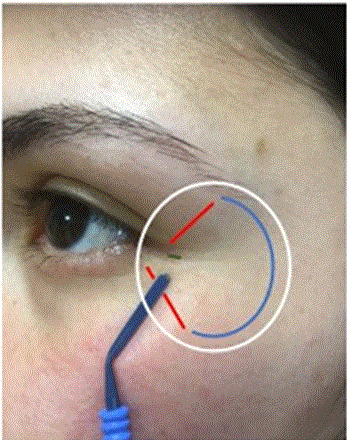
Figure 2: Marked surgical area. White: outline of undermined area. Red: myolisis. Blue: neurolysis. Green: incision.
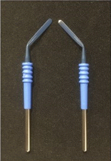
Figure 3: Left: Original coated angle electrode. Right: The electrode with unshielded tip.
All patients expressed satisfaction with the result at their 9-month postoperative check-up, including the patient with benign essential blepharospasm whose final check-up was 14 months after the procedure (Figure 4).
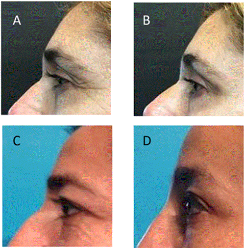
Figure 4: Before PEMN (A,C).Nine months after PEMN (B,D).
Electromyography showed an increase of threshold muscular activity in all patients. No paralysis of surrounding muscles was observed. There was no restriction of blinking and closure of eyelids. No enhancements were necessary.
Paresthesia in the surrounding area was referred to by 10 patients, 40 %, and disappeared at 3 months.
The appearance of facial wrinkles is multifactorial: genetic, racial, environmental, gender, aging, diet and habits such as smoking, but most important is the permanent contracture of the underlying muscles [7-9]. An often overlooked factor regarding the formation of crow’s feet is permanent squinting due to undiagnosed or uncorrected visual defects, especially in patients with myopia and/or astigmatism. It is always advisable for patients who squint to be assessed by an ophthalmologist, especially in the case of young people. Many efforts have been made to ameliorate facial wrinkles, such as: cosmetic care, topical medical agents, systemic agents, avoiding exogenous risk factors and invasive procedures [10]. Nowadays, the most acceptable method used to treat this stigmatic aging process is BT [11,12].
Surgical procedures imply OOM myectomy and neurotomy of the zygomatic branches [13-18]. The upper half of the OOM is innervated by one of the multiple rami of the temporal branch of the seventh cranial nerve (facial nerve), while the lower half by the zygomatic branch of the same cranial nerve [19,20]. The motor nerve fascicles travel under and perpendicular to the muscle fibers [21]. There are anastomoses between these terminal motor nerves and the sensory nerve fibers of the fifth cranial nerve (trigeminal nerve). Therefore, the zygomaticofacial and zygomaticotemporal nerves (sensory nerves), have a connection with the zygomatic nerve (motor nerve) [22,23], which would explain the paresthesia that some patients experienced in this study.
After referencing medical literature, the senior author, experienced in blepharospasm [24] and BT in the lateral canthal area, offered the new procedure, denominated PEMN, to former patients who had received treatment with BT. The operation was explained in detail and patients were given the possibility of further treatment if they were not satisfied with the result. Low intensity power was used on the first patients; they required BT touch-ups and were not included in this cohort. It was determined that the ideal power for this procedure is between 35 to 40 W.
One fear referred to by patients regarding this method was the impediment of eyelid closure. On this subject, McCord and Codner have stated that the buccal branch of the facial nerve commands the innervation of the inner canthal orbicularis and is responsible for blinking, closure, tone of the lower lid, and the pumping mechanism for the lacrimal apparatus [25].
Spontaneous nerve damage regeneration takes at least 3 to 4 months [26,27], which lead the authors to defer assessment of the result of the procedure until a minimum of 9 months had elapsed. A subsequent longer-term follow-up is also recommended.
The long-lasting results, efficacy and patient satisfaction demonstrated that the use of PEMN on the OOM could substitute BT for the attenuation of crow’s feet. Whereas the effect of BT lasts approximately four months, PEMN is an easy to perform technique and a permanent solution which does not normally require further treatment. In this study, repetition of PEMN was not necessary. Unlike neuromodulators which has an immediate visible effect, the results of PEMN can be observed once the post-operative swelling has subsided, generally after five days. With PEMN, a two-day downtime period is required.
Due to the excellent results and effectiveness of this procedure, the authors are considering its use in the treatment of glabellar wrinkles as well as its possible implementation in routine blepharoplasties. PEMN is also being evaluated as a possible replacement for BT to relax chewing muscles in the case of bruxism [28-30].
The author(s) declared no potential conflicts of interest with respect to the research, authorship, and/or publication of this article.
The author(s) received no financial support for the research, authorship, and/or publication of this article.
To Richard Cutler Allen, MD, whom I do not know personally but for whom I feel gratitude and admiration for his free online Oculoplastic Surgery Video Library. His more than 300 videos have been one of the best educational supports in my career.
Download Provisional PDF Here
Aritcle Type: SHORT COMMUNICATION
Citation: Schwember J, Madrid L, Hernández A, Leiva F, Kurtzig A (2022) Percutaneous Electromyoneurolysis of the Orbicularis Oculi Muscle: A Permanent Substitute of Botulinum Toxinin Crow’s Feet. J Clin Cosmet Dermatol 6(1): dx.doi.org/10.16966/2576-2826.170
Copyright: © 2022 Schwember J, et al. This is an open-access article distributed under the terms of the Creative Commons Attribution License, which permits unrestricted use, distribution, and reproduction in any medium, provided the original author and source are credited.
Publication history:
All Sci Forschen Journals are Open Access