Figure 1: Follicular Hyperkeratotic papules


Al-Mahmoud BE* Almaslamani HA Al Hayki NA
Department of Dermatology, Rumailah Hospital, Hamad Medical Corporation, Qatar*Corresponding author: Al-Mahmoud BE, Department of Dermatology, Rumailah Hospital, Hamad Medical Corporation, Qatar, Tel: +974 55866615; E-mail: Badria.6094@gmail.com
Eruptive Vellus Hair Cyst (EVHC) is a term proposed and published for the first time by Esterly et al. in 1977. EVHC is a benign disease, and its exact cause is unknown. Some consider it to be a hamartomatous differentiation toward Vellus hair follicles while others believe it is a rare developmental abnormality of vellus hair follicles. Any area of the skin surface may be affected with this condition. As proposed, the distribution of the lesions is related to the condensation of the pilosebaceous units in the skin. It can be sporadic or inherited as an autosomal dominant trait. The histologic feature of EVHC is monomorphic; the diagnostic hallmark of EVHC is multiple fragmented vellus hair shafts within the keratinous cyst in the middle dermis. The combination of EVHC with different pilosebaceous unit cystic differentiations and their incorporation with different clinical skin conditions and their presence in various genetic, endocrinal, and neurological conditions have been documented in the literature.
Though the prevalence EVHC in one study was 1.6%, EVHC is not a rare disorder as it appears to be, but its frequency is most probably underestimated due to the paucity of symptoms. In addition to the variability of its clinical features, it has a cosmetic disability, which is the chief concern of most patients. In 25% of EVHC patients, lesions regress spontaneously after transepidermal elimination or after the development of a foreign body granuloma by degradation of the cyst walls; however, treatment of persistent lesions is often challenging, with disappointing results.
Eruptive vellus hair cysts; Hyperpigmented monomorphic papular eruption; Cystic lesion; Rice grain-like lesions
Eruptive vellus hair cyst (EVHC) is a term proposed and published for the first time by Esterly et al. [1] in 1977; describing hyperpigmented monomorphic papular eruption in two children. Since then, many publications have reported EVHC cases with a broad range of clinical features, such as the cystic lesion, rice grain-like lesions, yellow brown and yellow red or blue papules, papulocystic, and as bluish-gray facial discoloration. EVHC may also appear as persistent acniform and even comedon-like lesions. The lesions can be single as a giant solitary lesion or multiple, localized or generalized, follicular and non-follicular in addition to umbilicated and non-umbilicated.
Though the chest and extremities are the most common sites of involvement in EVHC, any area of the skin surface can be affected [2], including the eyelid [3] and labia majora [4]. As proposed; the distribution of lesions was related to the condensation of the pilosebaceous units in the affected area [5]. The development of lesions is usually progressive, but not necessarily eruptive, contrary to what their name suggests.
EVHC can be sporadic and often appears without any antecedent trauma or triggering factor in the first and second decades of life [6]. An interesting subgroup of patients was reported to have had middle-age onset, meaning after the age of 35 [7]. The hereditary nature of this condition was discovered in 1980 [8,9]. Several reports have noted familial cases [10] and cased in twins [11]. The condition is inherited as an autosomal dominant trait, with lesions appearing at birth or in early infancy. EVHC has no racial predilection, and males and females are equally affected [8].
Unlike the clinical polymorphism of EVHC lesions, the histologic feature of EVHC is monomorphic; the diagnostic hallmark of EVHC is a keratinous cyst in the middle dermis, lined by a few layers of epithelial cells with rudimentary hair bulb-like structures. The cysts contain multiple fragmented vellus hair shafts and laminated horny material [12]. The immunohistochemical of the lesion revealed expressing antigens of the normal hair follicle infundibulum, such as keratins 1/10, calretinin, and p63, along with a reduced Ki67 proliferation index in the cyst wall [13]. The dermoscopic findings of EVHC include non-melanocytic lesions, welldefined, whitish to yellow, with circular structures and erythematous brownish halos. A central gray and blue color points were observed because of the presence of melanin within the pigmented hair shaft in the cyst [14].
EVHC is a benign disease. The exact cause is unknown. Some consider it to be a hamartomatous differentiation toward vellus hair follicles while others believe it is a hamartomatous growth of vellus hair follicles, predisposing them to occlusions at the level of the infundibulum, retention of hair, cystic dilatation of proximal part of the follicles, and secondary atrophy of the hair bulbs [15].
EVHC is part of pilosebaceous cyst spectrum originating in the pilosebaceous duct. The term “hybrid cysts” has lately been coined to describe the existence of pilosebaceous unit differentiation in the same cysts [16]. Multiple pilosebaceous cysts referred to the presence of more than one pilosebaceous cysts in the same pathologic process of EVHC, most commonly steatocystoma multiplex [13], syringoma, epidermoid, and trichilemmal cysts [17]. The diagnosis and the differentiation are established on histopathologic examination [18].
Though the prevalence EVHC in one study was 1.6% [19], EVHC is not as rare of a disorder as it appears. Its frequency is most probably underestimated due to the paucity of symptoms in addition to the variability of its clinical features and its incorporation with different clinical skin conditions such as acne vulgaris and keratosis pilaris. It creates a cosmetic disability, which is the chief concern of most patients. For 25% of VHC patients, lesions regress spontaneously after transepidermal elimination or after the development of a foreign body granuloma by degradation of the cyst walls [20]; however, the treatment of persistent lesions is often challenging with disappointing results [21].
When Esterly et al. [1] reported and proposed the name “Eruptive villus hair cyst” for the first time in 1977, they noticed spontaneous involution of the eruption in one of the two cases they reported. Spontaneous regression within months to years without treatment has been reported thereafter, but the natural history was not well defined [12]. Bovenmyer DA [22] reported EVHC case with umbilicated and non-umbilicated lesions. Histopathologically, he found communication to the surface of the skin with released vellus hair from the pores of the umbilicated lesions. The opening of the cysts to the epidermis was also detected by dermoscopy [23].
Herein, we describe the clinical details of two cases selected from my daily clinic in the Department of Dermatology & Venereology, Hamad Medical Corporation in Qatar, to clarify the exiting of the eruptive vellus hair cysts in the vicinity of different skin diseases and describe the diagnostic signs that direct my attention to the EVHC lesions.
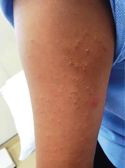
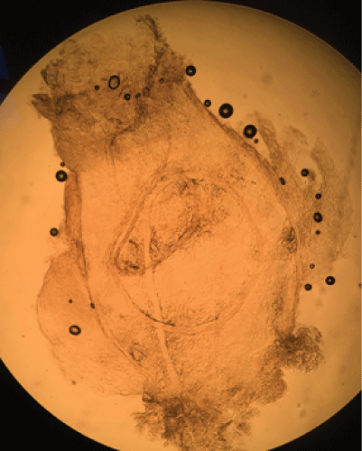
A five-year-old male presented with multiple, asymptomatic skin lesions on both upper arms, of 3 months duration. The child used to have a good general condition with positive family history of atopy. Physical examination revealed dry skin in general with discrete, symmetrically distributed follicular hyperkeratotic papules, a few of them with tiny white scales protruding from central follicular openings (Figure 1) on the lateral aspect of both upper arms. The condition was diagnosed as a case of keratosis pilaris. When the white scale pulled out non-surgically with non-toothed forceps and examined microscopically in potassium hydroxide preparation, shows a vellus hair cyst within an intact cyst (Figure 2).

Figure 1: Follicular Hyperkeratotic papules

Figure 2: Vellus hair cyst within an intact cyst
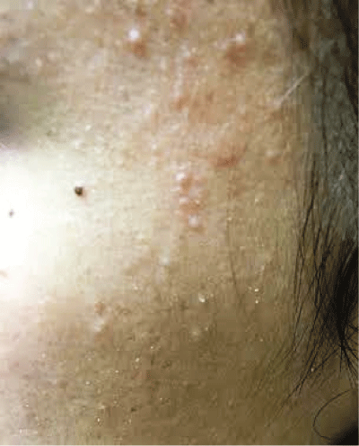
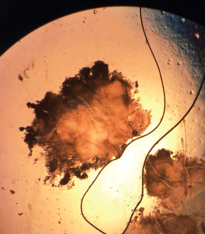
An 18-year-old female patient was presented with an asymptomatic acneiform eruption of face and neck with some lesions on the chest of 3 years duration. Several modalities of acne treatments have been used, topical and systemic, with no improvement. A skin examination showed inflammatory papules with black and white heads. In the same distribution, a few discrete lesions show follicular hyperkeratosis with the central protrusion of a little white scale (Figure 3). A vellus hair within intact cyst is shown with non-surgical pull out of the scale with non-toothed forceps and examined microscopically in a potassium hydroxide preparation (Figure 4).
.
Figure 3: Discrete lesions showing follicular hyperkeratosis

Figure 4: A vellus hair within intact cyst
EVHC are benign lesions of the pilosebaceous duct. They are inherited as autosomal dominant traits. In acquired and sporadic cases, we currently classify patients into two groups. First, we have primary idiopathic EVHC where the lesions arise spontaneously with no associated cutaneous or systemic condition. The secondary EVHC usually associate with other pilosebaceous cysts and different inflammatory skin conditions. They are also found in association with various medical, immunologic, genetic, and metabolic conditions.
The pathogenesis of EVHC is unknown. A theory of the developmental hamartoma of vellus hair follicle tissue has been postulated. Caused by a mutation in the keratin gene in the inheritance cases, it has an earlier onset compared with acquired cases. In a case of primary idiopathic EVHC, a local defect(s) can be responsible for the hamartoma changes in vellus hair follicle tissue. The pathogenesis is still obscure but may involve cytokines, stress proteins, or adhesion molecules. However, the actual stimulus, which is selective in inducing the vellus hair cyst rather than other pilosebaceous hamartomas lesions to form, is unknown. It is likely that secondary EVHC associated with an inflammatory skin condition represents a non-specific epithelial reaction pattern to a variety of different provoking stimuli. The faulty follicular degeneration associated with genodermatosis conditions may induce a sequestered hamartomatous overgrowth of the pilosebaceous epithelial cells, leading to vellus hair follicle occlusion in the infundibular level and cystic dilatation of the proximal part of the hair follicle with retention of keratin and hair. This theory probably explains the hamartoma developmental part of the pathogenesis story.
All reported cases of EVHC, whether primary or secondary, has monomorphus histologic picture of vellus hair cysts; Intra-dermal cysts, lined by epidermal-type epithelium and containing vellus hair and keratin. In spite of the keratin expression and immunohistochemical studies, it is hard to trace the original parent cells of these cysts and how they arise in the literature. There were no reported serial section studies for this condition. Possibly because of that, it was impossible to describe the proper pathogenesis, and the story of the developmental hamartoma has survived.
Due to its resemblance to variable similar pathologic conditions, the clinical presentation does not usually diagnose EVHC [24]. Diagnosis usually is confirmed by Punch biopsy and histological examination, or either puncturing the skin with the blood-collecting needle to aspirate cystic content or extracting the cyst out by dissecting forceps for histopathology or microscopic potassium hydroxide preparation examination. Nevertheless, these procedures can be both distressing and painful and may leave an unpleasant scar. In the present cases, we used an alternative method to support the clinical diagnosis; we introduce an efficient and a successful fast track nonsurgical technique for in-office diagnosis of EVHC. We suggest this method as the practical diagnostic tool in the diagnosis for this condition.
The current view of EVHC is that it is not a disease entity, but it can be principally considered as a reactive process often associated with different cutaneous pathologic conditions. On the other hand, the inconstant relationship of the polymorphic clinical pictures to the monomorphic pathologic findings, the delay to incorporate new pathophysiologic studies into clinical practice, and the lack of diagnostic techniques with which to objectively identify EVHC are complex nature of points that fulfill the definition of a phenomenon and provide many opportunities to address EVHC condition as phenomena rather than a disease. Exploring this condition and its natural context in depth is required in the future studies.
Download Provisional PDF Here
Aritcle Type: CASE SERIES
Citation: Al-Mahmoud BE, Almaslamani HA, Al Hayki NA (2018) Eruptive Vellus Hair Cysts: Is it a Disease or a Phenomenon? J Clin Cosmet Dermatol 2(2): dx.doi.org/10.16966/2576-2826.127
Copyright: © 2018 Al-Mahmoud BE, et al. This is an open-access article distributed under the terms of the Creative Commons Attribution License, which permits unrestricted use, distribution, and reproduction in any medium, provided the original author and source are credited.
Publication history:
All Sci Forschen Journals are Open Access