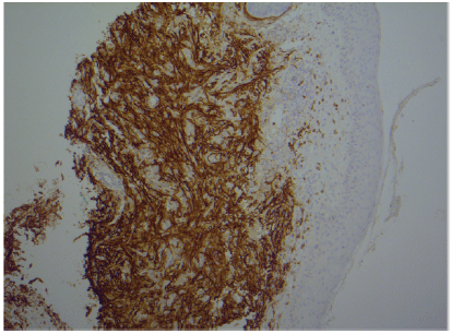
Figure 1: A figure confirming CD 10 positivity
Prativa SA Jayasekera1* Stuart A Osborne2 James AA Langtry3
1Royal Victoria Infirmary, Newcastle upon Tyne BM Medicine MRCP DERM, England, UK*Corresponding author: Prativa SA Jayasekera, Royal Victoria Infirmary, Newcastle upon Tyne BM Medicine MRCP DERM, England, UK, Tel: 0191 233 6161; E-mail: prativaj@googlemail.com
Atypical fibroxanthoma (AFX) is the most common of the rare cutaneous malignancies. AFX usually involves sites of high cumulative ultraviolet exposure and is rarely located in the periocular region. We are aware of 7 case reports in the literature, of periocular AFX, one of which was treated with Mohs Micrographic Surgery (MMS). We report our experience of a patient with AFX of the left medial canthus treated by MMS and review the literature of this rare tumour in the periocular region.
Atypical fibroxanthoma; Micrographic Surgery
A 55 year old woman with skin type II/III presented with a 10 month history of an enlarging, tender nodule affecting the left medial canthus. Her past medical history included asthma, hypothyroidism and atrial fibrillation and medications included levothyroxine. She had no past history of skin cancer. She went on sunny holidays as a child but rarely as an adult and had used sunbeds approximately 100 occasions in her lifetime. An incisional biopsy of the left medial canthal nodule confirmed AFX with immune histochemistry demonstrating CD10 positivity and cytokeratin negativity, figures 1, 2 and 3. Figure 4 shows a pre operative photograph. The tumour measured 13 × 13 mm and was excised by MMS. The tumour was cleared in one stage and two blocks (Figure 5). The final defect size was 23 × 19 mm and this was repaired by a myocutaneous cheek advancement flap. Two years post operatively she has had no sign of recurrence.
Figure 1: A figure confirming CD 10 positivity
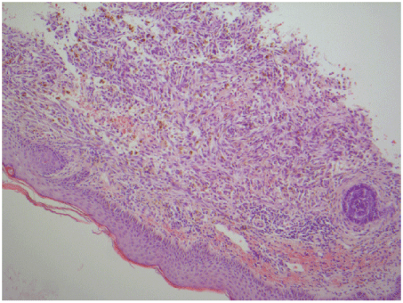
Figure 2: An H and E figure demonstrating marked cellularity and pleomorphism
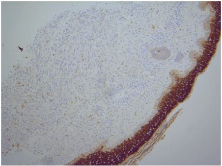
Figure 3: A figure showing negative cytokeratin.
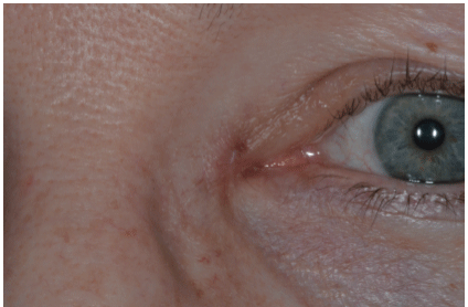
Figure 4: A medical photograph of a medical canthal nodule biopsied to be AFX
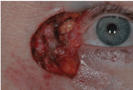
Figure 5: A photograph after tumour clearance with mohs micrographic surgery
AFX is a fibrohistiocytic tumour which commonly occurs on sun exposed skin. It was first described in 1951 by Helwig. AFX commonly presents as a rapidly growing, solitary nodule, which may ulcerate, be sessile or pedunculated. AFX commonly occurs on the head and neck region, but can occur on the trunk and limbs [1].
The clinical differential diagnoses include squamous cell carcinoma, amelanotic melanoma, pyogenic granuloma and cutaneous lymphoma.
Histologically, AFX is characterized by dermal proliferation of atypical spindle cells which are arranged in a haphazard or disordered pattern. The histological variants of AFX include granular cell, clear cell, desmoplastic, haemosiderotic, osteoclast-like giant cells, pigmented form and giant cell AFX [1,2].
It is important to distinguish AFX and pleomorphic dermal sarcoma (PDS), as the metastatic rate for PDS is in excess of 20% and rare for AFX.
AFX and PDS are histologically indistinguishable from each other. PDS
is diagnosed when the tumour extends beyond the dermis and represents a deeper variant of AFX. Therefore a superficial biopsy may lead to an
incorrect diagnosis of AFX. AFX and PDS may both demonstrate positive
immune histochemistry for vimentin, alpha anti-trypsin, CD68, CD99
and smooth muscle actin [2].
Risk factors for AFX include actinic damage secondary to ultraviolet radiation, immunosuppression, X-ray radiation, skin trauma and xeroderma pigmentosa [1-3].
Surgical excision is recommended for AFX, however there is no consensus on the surgical margin required. AFX rarely metastasises but local recurrence is not uncommon. Local recurrence after excision has been estimated to be between 2 and 20% [3]. Seavolt and McCall [4] described MMS for AFX in thirteen patients (ear n=3, scalp n=2, chest n=1, temple n=1, cheek n=2, leg n=1, upper lip n=1, jawline n=1, neck=1, eyebrow n=1) and at 2 years follow-up data was available for 6 patients, with no recurrence found.
In a retrospective study of 45 patients, 19 patients were treated with MMS and 26 patients were treated with wide local excision (WLE). MMS resulted in a lower recurrence rate of AFX compared to WLE. In patients treated with MMS there were no recurrences with a mean follow-up of 30 months and three recurrences in patients treated with WLE with a mean follow-up of 74 months. The margin of excision was documented in 16 out of 26 patients treated with WLE, ranging from 3 to 26 mm (mean 7 mm) [5].
One large study looking at AFX treated by MMS or WLE included 91 patients with 93 tumours. The most common location was the ear followed by the scalp. Information was available for 88 out of 93 tumours, of which 59 had MMS, 23 had WLE and 6 patients had other methods of excision (electro dissection, curettage and shave excision). 36 patients who had MMS required 1 stage to clear the tumour, 19 patients required two stages and 4 patients required more than two stages. The median margin required to clear the tumour with MMS was 4mm. There were no recurrences at follow-up (median 4.5 years) following MMS and there were two recurrences following WLE [6]. The current reported recurrence with MMS is between 0-6% and with wide local excision (WLE) 16% [6].
Periocular AFX is rare with seven cases reported in the literature, involving the medial canthus (n=1) [7] lower eyelid (n=4) [3,8-10], upper eyelid (n=1) [11] and lower eyelid and cheek (n=1) [12].
Table 1 presents all published cases of periocular AFX. The majority of the patients were male, with an average age of 63 years. The size of the tumour ranged from 6 × 6.5 mm to 60 × 60 mm (Table 1). Five patients underwent excision, one had MMS and the method of the tumour removal was not documented for one patient. There were no documented recurrences, at follow-up ranging from 6 months to 1 year.
| Author, Year of publication | Gender | Age | Location of tumour | Size in mm | Duration of tumour in weeks | Type of tumour removal | Recurrence | Follow-up in months |
| Haye C, 1981 | Female | 31 | Upper eyelid | 5 x 6 | 104 | Excision | Not documented | Not documented |
| J Boynton, 1989 | Female | 58 | Lower eyelid | 6 x 6.5 | 6 | Excision | Not documented | Not documented |
| C Rice, 1991 | Male | 82 | Lower eyelid and cheek | 60 x0 6 | Not documented | MMS | No | 12 |
| N Paramanthan, 2008 | Male | 61 | Lower eyelid | 5 | 8 | Excision | No | 12 |
| A Kilic,2008 | Female | 12 | Lower eyelid | 10 x 5 | Several months | Excision | No | 5 |
| U Kim, 2009 | Male | 78 | Lower eyelid | 60 x 50 | 156 | Excision | No | 6 |
| D Rahore, 2013 | Female | 90 | Medial canthus | 15 x 15 | Not documented | Excision | No | 12 |
Table 1: A table summarising published case reports of periocular AFX
Paramanathan et al. [9] reported a patient with recurrent AFX of the lower eyelid. The tumour was first removed with clear excision margins. Two months later it recurred within the scar site. The patient then underwent a second excision with a 5 mm margin, without recurrence at 12 months.
Rice et al. [12] reported a patient with a 30 mm diameter AFX, of the right lower eyelid and cheek treated by MMS. Tumour negative margins resulted in a defect size of 60 × 60 mm and there was no recurrence at 1 year follow-up.
Our case highlights the use of MMS for periocular AFX. MMS tumour clear margins allows a high level of certainty of tumour clearance with likely higher cure rate and tissue sparing which are critical in the surgical oncology of the periocular skin.
We advocate the use of MMS for AFX of the periocular skin in order to achieve tumour clear histological margins and the potential for tissue sparing.
Download Provisional PDF Here
Aritcle Type: Case Report
Citation: Jayasekera PSA, Osborne SA, Langtry JAA (2017) Atypical fibroxanthoma of the medial canthus treated by Mohs Micrographic Surgery. J Clin Cosmet Dermatol 1(3): doi http://dx.doi. org/10.16966/2576-2826.115
Copyright: © 2017 Jayasekera PSA, et al. This is an open-access article distributed under the terms of the Creative Commons Attribution License, which permits unrestricted use, distribution, and reproduction in any medium, provided the original author and source are credited.
Publication history:
All Sci Forschen Journals are Open Access