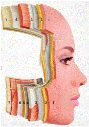
Figure 1: Face’s Stratigraphy: Epidermis, dermis, subcuneous, muscles, deep fat compartment and bone.


Patricia Fabrini1* Pedro Fabrini2 Natalia Centrone2
1Dermatologist MD Graduated in Medicine from University of Minas Gerais, Member of the Medical Staff at Hospital Israelita Albert Einstein, Brazil*Corresponding author: Patricia Fabrini, Dermatologist MD Graduated in Medicine from University of Minas Gerais, Member of the Medical Staff at Hospital Israelita Albert Einstein, Brazil, Phone: +5531984775497; E-mail: [email protected]
There are many approaches to the treatment of facial aging. However, all of them should be based on a comprehensive understanding of the face as a multi-layered anatomical structure. Facial aging is a dynamic process that affects every tissue layer, including the facial skeleton, retaining ligaments, deep and superficial fat compartments, the superficial musculoaponeurotic system (SMAS), muscles, subcutaneous tissue, dermis, and epidermis.
With aging, the facial skeleton undergoes progressive remodeling and bone resorption, reducing structural support for the overlying soft tissues. Facial retaining ligaments fibrous connective tissue structures that separate anatomical compartments and anchor the soft tissues to the underlying skeleton or deep fascia also undergo age-related changes, becoming less effective in maintaining facial support. In addition, fat compartments experience selective atrophy, hypertrophy, and redistribution, while the skin progressively loses collagen, elastin, and hydration.
The interaction of these changes across all anatomical layers results in the characteristic features of facial aging, including volume loss, tissue descent, skin laxity, and alterations in facial contour. Therefore, successful facial rejuvenation requires a thorough understanding of these agerelated modifications and a layer-by-layer treatment strategy rather than focusing on a single tissue plane (Figure 1).
Figure 1: Face’s Stratigraphy: Epidermis, dermis, subcuneous, muscles, deep fat compartment and bone.
The face is composed of multiple interconnected anatomical layers, including the skin, subcutaneous fat compartments, facial muscles, retaining ligaments, and the underlying facial skeleton [1].
A comprehensive understanding of the age-related changes occurring in each of these layers including the epidermis, dermis, superficial and deep fat compartments, muscles, retaining ligaments, and bone is essential for achieving natural, harmonious, and longlasting aesthetic outcomes.
These anatomical layers are interconnected by a complex network of collagen-rich retaining ligaments. These fibrous connective tissue structures provide structural support by anchoring the soft tissues to the underlying periosteum or deep fascia, maintaining facial architecture, compartmentalization, and mechanical stability. Importantly, retaining ligaments also undergo age-related structural and functional changes, contributing to facial ptosis and the characteristic features of facial aging [2,3].
The retaining ligaments divide the face into distinct anatomical compartments, serving as key structural boundaries that maintain facial architecture and regulate the position of the overlying soft tissues. Age-related attenuation of these ligamentous structures, combined with selective volume loss of the deep fat compartments particularly those located posterior to the premasseteric ligament plays a pivotal role in the development of facial ptosis and the characteristic morphological changes associated with facial aging (Figures 2,3).
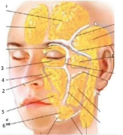
Figure 2: Role of retaining ligaments and deep fat compartments in facial aging.
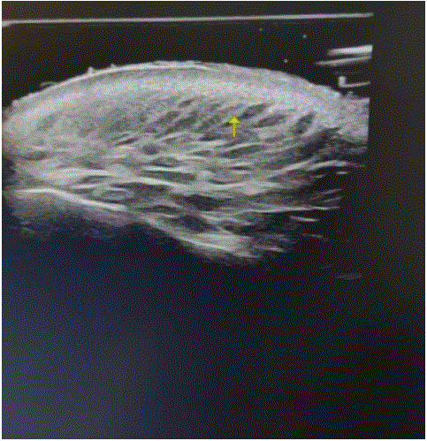
Figure 3: Ultrasound image of ligament connecting all the layers of the skin.
Treatment should begin with an appropriate skincare routine, which prepares the skin to better receive subsequent technologies and procedures.
Electroporation associated with bioactive substances such as PDRN (polideoxirribonucleic acid) promotes the restoration of damaged cells along the ligamentous network, leading to an immediate improvement in skin quality and appearance (Figure 4).
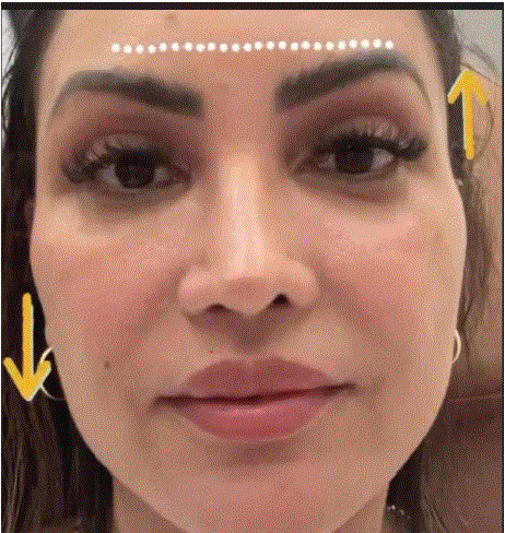
Figure 4: Stimulating ligaments can elevate all the layers of the skin.
When indicated, biostimulatory injectables (poly-l-lactic acid) can be placed beneath the retaining ligament plane to restore structural support and compensate for the progressive volume loss of the deep fat compartments that occurs with aging. This approach helps reestablish facial contours while preserving a natural appearance.
Finally, botulinum toxin plays a fundamental role in facial myomodulation by selectively reducing the downward pull of the depressor muscles in the lower third of the face. By restoring a more balanced muscular interplay, it contributes to improved facial support, softens the appearance of tissue descent, and enhances the overall rejuvenation outcome [4].
A comprehensive understanding of the anatomy and age-related changes affecting each facial layer allows for a personalized, layer-bylayer treatment strategy. By addressing the skin, retaining ligaments, fat compartments, muscles, and underlying skeleton, we can effectively manage both intrinsic facial aging and the anatomical changes associated with significant weight loss while preserving natural facial expressions, harmony, and long-lasting aesthetic results (Figure 5).
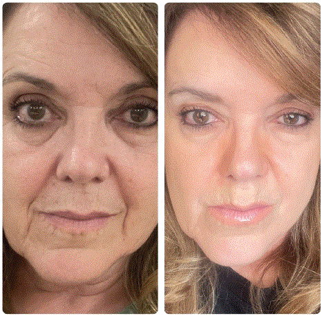
Figure 5: Before and after SKINCARE, eletroporation for ligament’s stimulation, miomodulation with toxin botulinic, and PLLA behind masseteric ligament.
Multiple anatomical and clinical studies identify the facial retaining ligaments as fundamental structures for maintaining facial support and guiding soft tissue repositioning during rejuvenation procedures. However, descriptions of the retaining ligament system vary considerably throughout the literature, reflecting differences in anatomical interpretation, dissection techniques, and terminology. Despite these variations, there is broad consensus that these ligamentous structures play a pivotal role in facial architecture, compartmentalization, and the aging process [5,6].
A thorough understanding of facial anatomy highlights the critical role of the retaining ligaments in maintaining facial support and underscores the importance of preserving and stimulating these structures as part of facial rejuvenation strategies.
Recognizing the dynamic continuity between the facial layers and the complex cellular crosstalk that integrates the skin, extracellular matrix, adipose tissue, muscles, ligaments, and bone supports a comprehensive, layer-by-layer treatment approach. Beginning with an optimized skincare regimen and addressing each anatomical layer according to its biological role allows clinicians to achieve more natural, harmonious, and long-lasting aesthetic outcomes.
Download Provisional PDF Here
Article Type: OPINION ARTICLE
Citation: Fabrini P, Fabrini P, Centrone N (2026) Facial Anatomization-A New Concept Based on Skin’s Stratigraphy. J Clin Case Stu 11(2): dx.doi. org/10.16966/2471-4925.295
Copyright: © 2026 Fabrini P, et al. This is an open-access article distributed under the terms of the Creative Commons Attribution License, which permits unrestricted use, distribution, and reproduction in any medium, provided the original author and source are credited.
Publication history:
All Sci Forschen Journals are Open Access