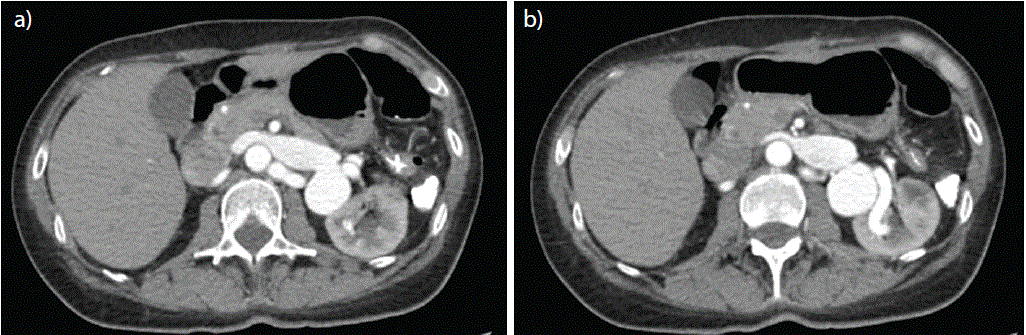
Figure 1: a) Left renal vein dilated, distal hiliar saccular aneurysm. b) Normal left renal artery diameter. Left renal vein filled by contrast in an arterial phase.


Jorge Adalberto Márquez1 Laura Valentina Llanos Luengas2* Veronica Saza Caicedo3
1Vascular and Endovascular Surgeon, Sociedad de Cirugía de Bogotá Hospital de San José, Bogotá, Colombia*Corresponding author: Laura Valentina Llanos Luengas, Vascular Surgery Fellowship Program, Sociedad de Cirugía de Bogotá Hospital de San José, Bogotá, Colombia, Phone: +57 3208009966; E-mail: [email protected]
Renal arteriovenous fistula (RAVF) is a rare condition, with a reported incidence of less than 0.04% [1], and is often associated with an asymptomatic presentation. However, some reported symptoms are macroscopic hematuria accompanied by abdominal pain, and in severe cases, it may be associated with heart failure [2]. The development of renal vein aneurysms has been associated with the presence of RAVFs [3], with the risk of rupture representing the most significant complication. A 60-year-old woman case is reported with an incidental finding on abdominal contrast computed tomography scan of a left RAVF, confirmed by angiography. The patient was completely asymptomatic and there was no interventional indication.
Renal vein aneurysm; Renal arteriovenous fistula; Vein aneurysm; Visceral aneurysm
A 60-year-old woman was sent to the emergency department from a neurology appointment with a 6-month history of lower limb weakness, progressive, limited standing and walking with right lateropulsion associated with frequent falls. She had a medical history of dyslipidemia, and had been on statin therapy for 6 months. She denied abdominal pain, hematuria, history of abdominal trauma or urinary tract manipulation, smoking, alcohol and the only prior surgical intervention was #4 uterine curettage due to abnormal uterine bleeding. She had not any genetic conditions associated with connective tissue weakening. Patients' CPK levels were markedly elevated (14356 U/L), electromyography revealed a myopathic pattern predominantly affecting the distal lower extremities leading to a probable diagnosis of immune-mediated necrotizing myopathy. Considering rapidly progressive symptoms and an inflammatory pattern, the neurology team prescribed a 5 days glucocorticoid therapy with a significant improvement of symptoms. No previous use of corticoids. No vital signs abnormalities were found, nor was marfanoid phenotype identified. On examination, a continuous bruit and important palpable thrill was felt in the left lumbar region, the inferior limb weakness was symmetrical. Uroanalysis found hematuria. A contrast computed tomography scan of the abdomen showed a saccular aneurysmal dilation of the left renal vein extended toward the renal hilum, measuring up to 48 × 40 × 48 mm, with multifocal fistulous communication with branches of the renal artery at the hilum (Figures 1a,1b). Aortogram with selective angiography of the left renal artery confirmed the diagnosis of an arteriovenous fistula in the distal third of the left renal artery (Figure 2). The patient was asymptomatic, there were no signs of cardiac failure, we didn’t find any interventional indication and she also denied any procedure. Patients' inferior limb weakness improved and was discharged without any symptoms, statins were suspended.
Figure 1: a) Left renal vein dilated, distal hiliar saccular aneurysm. b) Normal left renal artery diameter. Left renal vein filled by contrast in an arterial phase.
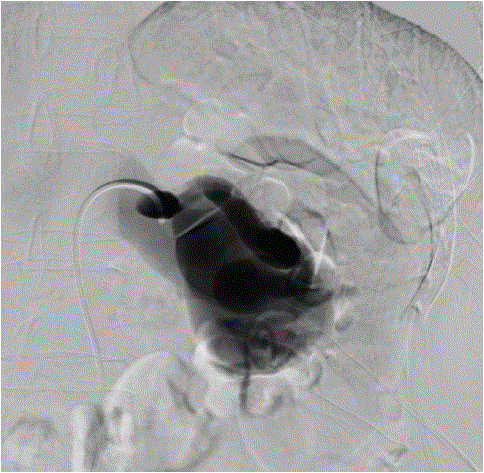
Figure 2: Arteriovenous fistula in the distal third of the left renal artery.
Renal arteriovenous fistula are relatively rare and most commonly acquired from blunt or penetrating abdominal trauma or invasive urinary procedures [4]. The diagnosis interval between the trauma and the identification of a RAVF can vary widely, with cases reported even decades later [5] due to an asymptomatic presentation. Angiography remains the best diagnosis and treatment method. Certain cases may require embolization or occlusion of the renal vein. In more severe presentations, particularly in distal or hilar fistulas, nephrectomy may be necessary. In asymptomatic patients, without cardiac, urinary or lumbar symptoms, intervention is generally not indicated, however, close and regular monitoring is recommended.
No conflicts of interest.
Written informed consent was obtained from the patient prior to publication.
Download Provisional PDF Here
Article Type: CASE REPORT
Citation: Márquez JA, Luengas LVL, Caicedo VS (2026) Incidental Renal Arteriovenous Fistula in an Asymptomatic Patient: A Rare Vascular Anomaly. J Clin Case Stu 11(2): dx.doi.org/10.16966/2471-4925.292
Copyright: © 2026 Márquez JA, et al. This is an open-access article distributed under the terms of the Creative Commons Attribution License, which permits unrestricted use, distribution, and reproduction in any medium, provided the original author and source are credited.
Publication history:
All Sci Forschen Journals are Open Access