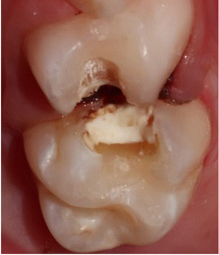
Figure 1: Preparation for inlays and Crown restorations.


Leonardo Rubio Arguello1* Brandon Palacios Irygoyen2
1Faculty Intercontinental University, Private Practice, Mexico City, Mexico*Corresponding author: Leonardo Rubio Arguello, Faculty Intercontinental University, Private Practice, Mexico City, Mexico, E-mail: [email protected]
Purpose: To describe a novel single-visit digital workflow for chairside fabrication of 3D-printed inlays, onlays and crowns in primary teeth and to report preliminary clinical outcomes.
Methods: Pediatric patients presenting with extensive carious lesions in primary teeth with or without signs of irreversible pulpal pathology were treated using a fully digital protocol. After conservative preparation, intraoral scanning and design using a CAD software, the restorations were printed chairside with a 3D printer using a biocompatible resin. Restorations were post-cleaned, cured and cemented with a resin cement during the same appointment. Clinical performance and short-term outcomes were evaluated.
Results: All restorations (n=11) were successfully delivered in a single visit with satisfactory marginal adaptation, esthetics and occlusal relation. No postoperative sensibility and debonding as well as two marginal small fractures that were easily repaired with composite was observed after a follow-up period of 24 months.
Conclusions: Single-visit chairside 3D-printed esthetic indirect restorations in primary teeth represent a feasible and promising minimally invasive alternative in selected cases. Further longitudinal studies are needed to determine long-term clinical performance.
Digital workflow; CAD/CAM; 3D-printing; Esthetic restorations; Pediatric dentistry; Primary molars; Chairside indirect restorations
A digital workflow in Dentistry encompass processes to obtain 3-D models of the oral cavity in an effort to substitute the analog workflows.
In Pediatric Dentistry we haven’t done procedures like indirect esthetic crowns, inlays and onlays restorations between other type of procedures because it implies the use of analog workflows that are more expensive, time consuming and unpractical for treating our pediatric patients.
With the advent of CAD-CAM (Computer Aided DesignComputer Aided Manufacturing) systems the use of a digital workflow in Dentistry have become increasingly popular with multiple applications in the different areas of Dentistry [1].
One of these CAD-CAM systems is the three-dimensional (3-D) printing, a state of the art technology that can be more affordable and easy to implement in a Pediatric Dentistry practice.
The continous improvement of intraoral scanning, CAD software, 3D printers and physical and biological properties of materials (resins, plastics, metals and ceramics) allows for increased accuracy and speeds the adoption of this developing technology with great impact in different fields of Dentistry [2] including Pediatric Dentistry where we can deliver more convenient cost effective, time efficient and accurate treatments to our patients. Compared to subtractive milling, 3D printing allows for lower production costs, reduced material waste and greater design flexibility [3,4].
The purpose of this article is to describe a novel chairside digital protocol for the fabrication of 3D printed inlays, onlays and crowns in primary teeth and to present preliminary clinical outcomes through an illustrated case series.
Pediatric patients presenting with extensive carious lesions where direct composite restoration was deemed biomechanically insufficient in primary molars and first permanent molars with Molar Incisor Hipomineralization (MIH), with or without signs of pulpal involvement which in tshis case will require a pulpal treatment prior the fabrication of the restoration, with sufficient remaining tooth structure to allow adhesive restoration and with two thirds of root length. Written informed consent was obtained from parents or legal guardians prior to treatment.
All procedures were performed under local anesthesia, rubber dam isolation and selective caries removal. For crowns restorations the reduction was similar as the stainless steel crowns reduction with slight differences like more occlusal reduction (1.5mm) and a feather edge enamel margin.
For inlays and onlays the cuspal reduction was between 1 and 1.5 mm, rounded internal line angles and smooth margins with a slightly beveled enamel margin (Figure 1).
Figure 1: Preparation for inlays and Crown restorations.
Following preparation, retraction cords were used (#0 and #00 Ultrapack, Ultradent Products, South Jordan, Utah, USA) and both the prepared and the antagonist arch were scanned as well as the bite relation (3600 Carestream Scanner, Carestream, Rochester, USA) and captured digitally (Figure 2).
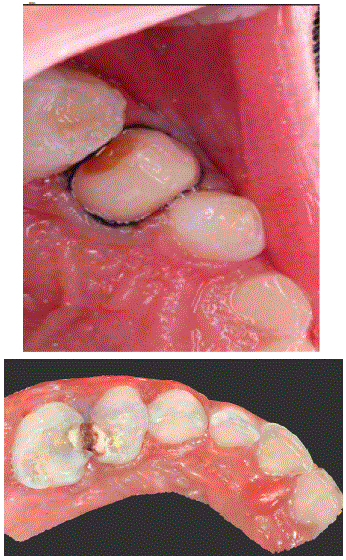
Figure 2: Scanning of the preparations.
Restorations were designed using a CAD software (Exocad GmbH, Darmastadt Germany). Occlusal morphology was customized to maintain functional occlusion, the proximal contacts were adjusted to ensure a passive insertion, and the cement space was set between 50- 100 microns, the orientation was between 120-150º to reduce support contact margins and the placement of the supports was limited to non-critical surfaces according to the manufacturer recomendations (Figure 3).
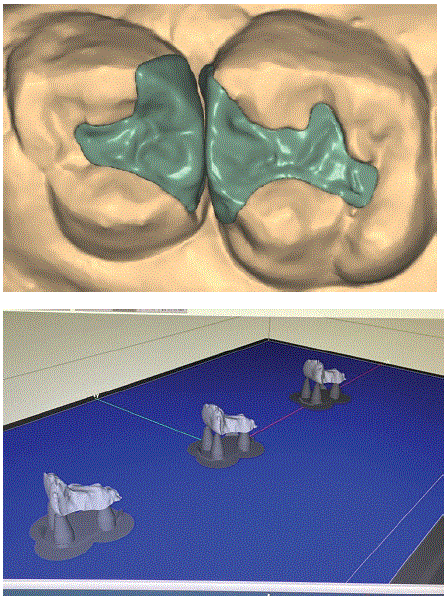
Figure 3: Exocad designs of two inlays and nesting in the printer software.
The design was exported in STL format and processed in a digital light processing (DLP) 3D printer software to fabricate the restorations chairside using a high resolution 3D printer (Cara Print, Kulzer, Hanau, Germany) (Figure 3). The printable resins used were biocompatible, light curable and indicated for long-term intraoral use (Dima Print Crown and bridge resins (Kulzer, Hanau Germany) (Figure 4).
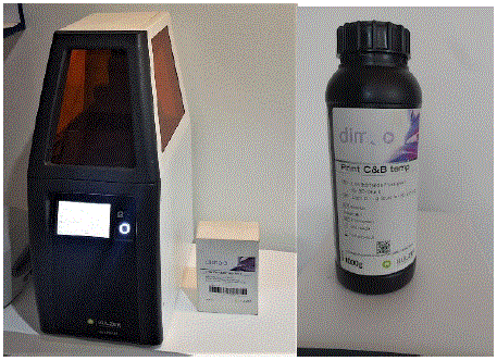
Figure 4: 3D-printer and printable resin.
After printing, restorations were cleaned in isopropyl alcohol in a Ultrasonic cleaner between 60-80º C (Anelsam, Mexico City, Mexico) twice for 3 minutes each to remove uncured resin (Figure 5) and subsequently post-cured for 5 minutes in a light-curing unit (HiLite power 3D, Kulzer, Hanau Germany) to achieve optimal mechanical properties (Figure 6).
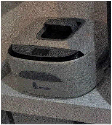
Figure 5: Cleaning in a ultrasonic cleaner with isopropyl alcohol.
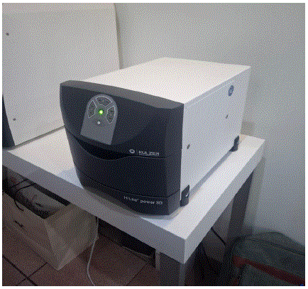
Figure 6: Post-cured in a light curing unit.
The supporting structures were carefully cut with pliers and surfaces were finished and polished using rotary polishing systems (Figure 7).
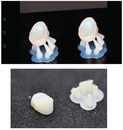
Figure 7: Restorations after the curing process and cut down of the supports.
The inner surfaces of the restorations were etched with 9% hydrofluoric acid (Ultradent Products, South Jordan, Utah, USA) for one minute, received a layer of silane followed by air drying and two layers of a universal adhesive system (3M, ESPE, Minneapolis, USA) followed each one by an air flow (1-2 seconds) to volatilize the solvent and finally light cured according to manufacturer guidelines with a LED photocured lamp (Coltene, Whaledent Inc, Cuyahoga Falls, Ohio, USA).
A total of twelve indirect restorations fabricated using the described chairside 3-D printing workflow were placed in eleven primary molars and one MIH first permanent molar. The restorations included four inlays, seven full coverage crowns and one onlay in 11 primary molars and one MIH first permanent molar with extensive affected surfaces. Clinical characteristics and outcomes are summarized in table 1.
| Case | Age (years) | Tooth (FDI) | Tooth type | Restoration type | Clinical outcome at 24 months |
| 1 | 4 | 54 | Maxillary first primary molar | Inlay | Successful |
| 2 | 5 | 64 | Maxillary first primary molar | Inlay | Successful |
| 3 | 6 | 55 | Maxillary second primary molar | Crown | Successful |
| 4 | 7 | 65 | Maxillary second primary molar | Crown | Successful |
| 5 | 5 | 75 | Mandibular second primary molar | Crown | Successful |
| 6 | 6 | 85 | Mandibular second primary molar | Crown | Successful |
| 7 | 4 | 74 | Mandibular first primary molar | Inlay | Minor marginal fracture |
| 8 | 5 | 84 | Mandibular first primary molar | Inlay | Successful |
| 9 | 6 | 75 | Mandibular second primary molar | Crown | Minor marginal fracture |
| 10 | 7 | 65 | Maxillary second primary molar | Crown | Succesful |
| 11 | 6 | 54 | Maxillary first primary molar | Crown | Successful |
Table 1: Case-by-case clinical outcomes of chairside 3D-printed restorations in primary molars (24‑month follow-up).
All restorations were successfully fabricated and delivered within a single clinical appointment and the digital workflow (Figure 8) was completed chairside without the need for provisional restorations and laboratory involvement.
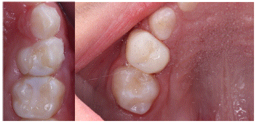
Figure 8: Restorations after cementation.
All restorations were clinically evaluated during a follow-up period of 24 months. The evaluation included restoration survival, marginal integrity, presence of fractures, retention, secondary caries and overall esthetic performance.
Out of the 11 restorations placed, all of them remained functional at the two year follow-up with a survival ratio of 100%. Minor complications were recorded in two restorations (1 inlay, 1crown) where small marginal fractures were detected and easily repaired with composite resin. There were not cases of debonding or secondary caries, adequate oclusal stability and the esthetic performance was satisfactory. The clinical performance of the restorations is summarized in table 1.
The findings in this study suggest that chairside 3D-printed restorations may represent a promising restorative alternative for primary teeth affected with extensive caries or developmental defects. The possibility of completing the entire digital workflow in a single visit offers additional advantages in Pediatric Dentistry where treatment time, patient cooperation and procedural simplicity are critical factors for clinical success [5].
Indirect restorations in primary molars have traditionally been associated with stainless steel crowns that present esthetic limitations which has led to the development of alternative options such as zirconia crowns and other esthetic full-coverage restorations, all of these are standard pre-made in a variety of sizes with the correct size being chosen to fit the trimmed down tooth. Some of these restorations also require more circumferential tooth reduction, are very difficult to adapt in cases of crowding, provide inaccurate marginal adaptation that is compensate with the cementing agent and inaccurate oclusal relations with the antagonists teeth [6]. A recent study concluded that prefabricated zirconia crowns had a higher mean value of marginal gap than 3-D printed resin crowns [7].
The two minor marginal fractures observed in the present study did not compromise the functionality or retention of the restorations and didn’t require replacements because were easily repaired. These findings are related with the material physical properties of currently available 3D-printing resins that present lower fracture resistance compared with other materials like ceramic milled or metals. Nevertheless, this type of resins are continously improving and at the present time we have printing resins with acceptable compressive and wear resistance that can resist the children masticatory forces.
With this digital chairside workflow one important advantage is the elimination of laboratory steps allowing clinicians to do the whole procedure during the same appointment that reduce treatment time and the need for multiple visits so now, pediatric dentists can do exact indirect restorations in primary molars, something that was imposible to do in the past.
The initial costs of 3D printing, cleaning, post-curing equipment and the resin are substantial, however, once the manufacturing process is established with clear design files in the STL format, in the long term this restorations are anticipated to be more cost- effective than certain prefabricated esthetic crowns [8].
It is important to say that this digital workflow opens new horizons in the Pediatric Dentistry practice because in addition of the restorations described in this article there are many other procedures that can be perform in one single visit at the dental office like Maryland Bridges in case of avulsed primary teeth, dental prostheses, non-nutritive habit devices, space maintainers, aligners, retainers, splints, and for sure in the near future many other more reducing time and costs of production [9] (Figures 9,10).
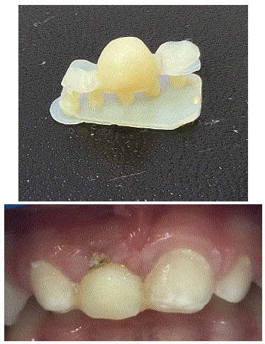
Figure 9: Maryland Bridge for an avulsed primary upper right incisor.
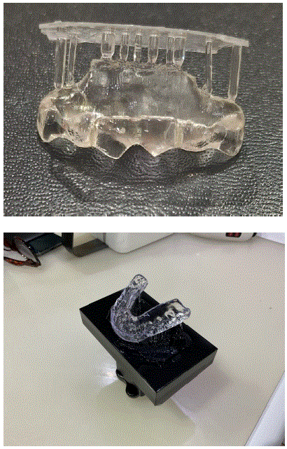
Figure 10: Tongue thrust habit device and splint.
Despite the promising clinical outcomes, this study presents several limitations, the relatively small sample size and the case series design limit the generalizability of the findings. Future studies with larger simples, controlled clinical trials and longer follow-up periods are necessary to confirm the reliability and long-term stability of chairside 3D-printed restorations in Pediatric Dentistry.
Within the limitations of this case series, chairside single visit 3D-printed esthetic restorations demonstrated favorable clinical performance in primary molars over a 24-month follow-up period.
All restorations remained functional with only minor fractures in two cases that required minimal intervention.
These restorations provided adequate esthetic outcomes, marginal adaptation and fracture resistance with high levels of patient and parent satisfaction.
The use of digital chairside workflow allowed the manufacturing and placement of customized indirect restorations in one single visit that is particularly advantageous in Pediatric Dentistry.
Three dimensional (3D) printing can provide more personalized, precised and accurate fabrication of dental restorations, reducing time and improving patient satisfaction.
As digital technologies become more prevalent in Pediatric Dentistry they can lead to develop many other kind of treatments, reducing costs and improving overall health and quality of life for our pediatric patients.
Download Provisional PDF Here
Article Type: CASE SERIES
Citation: Rubio L, Palacios B (2026) Single Visit Chairside 3D-Printed Esthetic Inlays, Onlays, Crowns and Other Applications in Pediatric Patients: A Clinical Innovation and Case Series. J Clin Case Stu 11(1): dx.doi.org/10.16966/2471-4925.287
Copyright: © 2026 Rubio L, et al. This is an open-access article distributed under the terms of the Creative Commons Attribution License, which permits unrestricted use, distribution, and reproduction in any medium, provided the original author and source are credited.
Publication history:
All Sci Forschen Journals are Open Access