Abstract
Objectives: Pneumocystis Jirovecii, a pathogenic fungus, causes severe interstitial pneumonia among immunocompromised patients. The transplant patients are predisposed to P. jirovecii pneumonia (PJP), most notably those under immunosuppressive therapy or with graft rejection. We describe here two cases of renal transplant cases with PJP and demonstrate the importance of molecular biology in the diagnosis and the prevalence of the DHPS gene mutation.
Material and methods: Diagnosis of Pneumocystis Jirovecii was made using conventional techniques: direct examination with staining GomoriGrocott modified by MUSTO (GG), May-Grünwald Giemsa (MGG), and Direct immunofluorescence (DIF) MONOFLUO™ Pneumocystis Jirovecii IFA Test Biorad. Molecular diagnosis was done with real-time PCR (Liferiver™ Pneumocystis Jirovecii®), and polymerase chain reaction-restriction fragment length polymorphism (PCR-RFLP).
Results: For the two samples, MGG, GG, and DIF staining did not allow the detection of a cystic or vegetative form. Real-time PCR reveals positive results for both samples for a quantity of DNA 103 copies per milliliter and 104 copies per milliliter. Amplification of the DHPS gene shows that no mutation has occurred in this gene. An improvement in the general condition of patients after 14 days of treatment with Sulfamethoxazole/ Trimethoprim was noted.
Conclusions: Our study shows that PCR is a faster and more sensitive technique than staining techniques. Currently, real-time PCR offers both a quantitative and qualitative approach with a very low detection threshold which makes it possible to differentiate between simple colonization and infection.
Keywords
Pneumocystis Jirovecii; Pneumonia; Real-time PCR; Renal transplant
Introduction
Pneumocystosis is an opportunistic mycosis caused by Pneumocystis jirovecii, a nubiquitous eukaryotic microorganism, classified among fungi in 1980 [1]. Up to 45 % of human immunodeficiency virus (HIV) patients suffer from Pneumocystis pneumonia (PJP). In addition, PJP cases in non-HIV-patients are increasing due to the use of glucocorticoids, chemotherapy, or other immunosuppressive drugs [1-3]. Furthermore, it is an increasing problem in transplanted patients: in the USA, up to 25 % of these patients suffer from a PJP [3].
The microbiological diagnosis of the PJP is usually based on microscopic demonstration of the fungus in the respiratory specimens by staining methods. The development of molecular biology techniques has significantly improved the diagnosis [4-6]. Molecular methods show more sensitivity and specificity compared to microbiological methods [7,8]. Furthermore, by quantitative PCR (qPCR), the quantity of the fungus in a specimen and even the status of the patient can be determinate [4,9,10].
The prophylaxis and the treatment of this disease are based on sulfonamides especially trimethoprim-sulfamethoxazole combination (TMP-SMX) or cotrimoxazole. As result, cases of resistance were selected with a mutation at the ‘dihydropteroate synthase (DHPS)’, the target of sulfonamides [11,12].
We describe here two cases of renal transplant patients with PJP and demonstrate the importance of molecular biology in the diagnosis and the prevalence of the DHPS gene mutation.
Methods
Case description
Case 1: A 53-year-old woman, suffering from renal failure caused by polycystic kidney disease, with-renal transplant in 2016 with chronic rejection under Mycophenolate Mofetil-tacrolimus-Prednisolone sodium metasulfobenzoate. She was admitted for an altered general condition consisting of asthenia, anorexia, fever of 39°C, and polypnea. Laboratory data showed C reactive protein (CRP) of 355mg/ml, Hb=9.2g/dl, WBC=7800/mm3, PLT=181000/mm3, urea=14.5mmol/l, creatinine=303µmol/l, K=4.2mmol/l, serum calcium=2.53 mmol/l. Chest X-Ray showed bilateral alveolar-interstitial syndrome. Serology for Candida spp and Aspergillus spp was negative; search for BK in the sputum was negative. Cytomegalovirus (CMV) PCR was negative. Microbiological investigation showed a urinary tract infection by Escherichia coli and a positive P. jirovecii real-time PCR. The patient was initially put on Tazocillin-Ciprofloxacin-Sulfamethoxazole/ Trimethoprim, then on Ceftazidime and Sulfamethoxazole/ Trimethoprim. General condition after one week with a second negative P. jirovecii PCR.
Case 2: A man, 62 years old with-kidney transplant since 2002. Graft biopsy puncture in 2018 showed idiopathic extra membranous glomerulonephritis which recurred when put on Cyclosporine and a high dose of corticosteroid. The clinical examination on admission noted a fever of 38°C and edema of the lower limbs. The chest X-Ray showed an alveolar-interstitial syndrome.
Serology for Candida spp and Aspergillus spp were negative. Search for BK in urine and sputum was negative. Blood and urine cultures were negative. CMV PCR was positive with 1136copies/mL. Also, PCR for P. jirovecii was positive. The patient was treated on Cymevan, Augmentin, and Sulfamethoxazole/Trimethoprim, with a reduction of the dose of immunosuppressive drugs.
The evolution was marked by improvement in the general condition after 14 days with a second PCR P. jirovecii negative.
Microbiological methods
Respiratory specimens (sputum for both patients) were centrifuged at 6000g for 10min. Part of the suspended pellet (about 200µl) was used for microscopic examination (MGG, GG stains) and 200µl was used for real-time PCR for detection and quantification of P. jirovecii DNA and PCR-RFLP for the detection of DHPS gene mutations in P. jirovecii strains.
Conventional techniques-microscopy
The MGG stain highlights intra-cystic bodies and/or trophozoites of P. jirovecii. Giemsa stains the nuclei of all Pneumocystis life cycle stages in purple-pink but does not stain the cell wall while GG stains the wall of cysts of P. jirovecii in black brown on a green background.
The DIF using the kit MONOFLUO™ Pneumocystis Jirovecii IFA Test (Biorad) allows highlighting the presence of pathogen (cyst or vegetative form) by immunological labeling. The attachment of the pathogen to the fluorescein-labeled antibody gives an apple-green coloring according to manufacturer’s instructions for use.
Molecular diagnosis
Real-time PCR using the Life river™ Pneumocystis Jirovecii® allows the detection and quantification of P. jirovecii DNA. PCR-RFLP was used for DHPS locus analyzes.
Results
For two samples, the MGG and GG staining did not allow the detection of a cystic or vegetative form. The DIF technique also remains negative. Real-time PCR reveals a positive result for two samples 103 copies per milliliter and 104 copies per milliliter respectively.
(Figure 1) showed the quantification of P. jirovecii DNA. (Figure 2) illustrates the amplification of the DHPS gene at 370bp with conventional PCR. PCR RFLP shows that no mutation has occurred in this gene for the two cases (Figure 3)
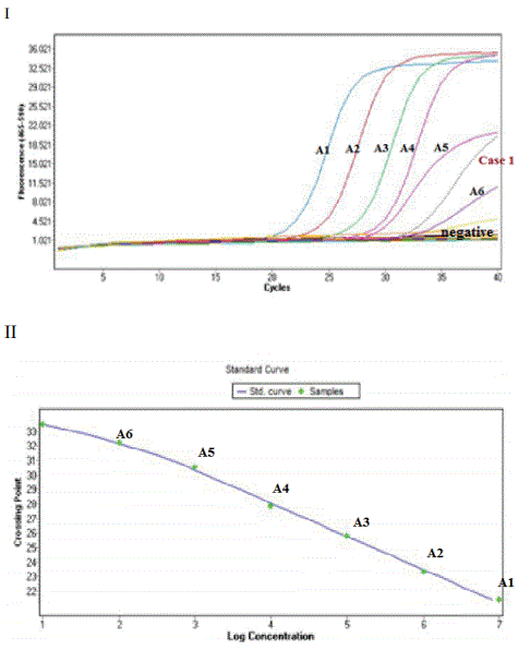
Figure 1: The curve of the quantification of PJ DNA by real-time PCR.
I: The amplification curves; A1 to A6: standards with a DNA concentration of 107 to 10 2 copies/µl; A1 (107), A2 (106), A3 (105), A4 (104), A5 (103), A6 (102), Case 1: the sample tested; Negative: negative control.
II: The standard curve constructed from the values of Cp (crossing point) versus the log DNA copy number/(range=107 to 102 copies/µl).
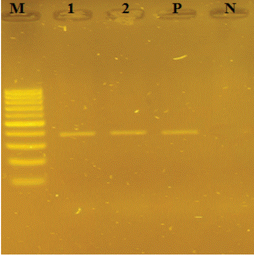
Figure 2: Agarose gel electrophoresis of conventionnal PCR products of P. jirovecii DHPS gene 370 pb.
Lanes: M (100 bp DNA size marker); 1 (Case 1); 2(Case 2); P (positive control); N (negative control).

Figure 3: Agarose gel electrophoresis of RFLP-PCR products of P.
jirovecii DHPS gene by accI and HaeIII.
Lanes: M (100 bp DNA size marker); 1and 2(Case 1 and Case2 aftre restriction by AccII); I and II (Case 1 and Case 2 after restriction by HaeIII).
An improvement in the general condition of patients after 14 days of treatment with Bactrim® was noted. Even real-time PCRs for P. jirovecii after this interval are negative.
Discussion
Currently, cases of pneumocystosis in HIV-infected patients tend to decrease due to the use of anti-Pneumocystis Jirovecii chemoprophylaxis [1,4,13]. Nevertheless, its incidence has dramatically increased over the last 15 years in patients with other predisposing immunodeficiencies [1], particularly transplant recipients [14-17]. In our study, we diagnosed the presence of P. jirovecii in two renal transplant patients, non-HIV. Magne, et al. reported 805 cases of PJP including 264 (33%) non-HIV, 86 had undergone a transplant among them 37 kidney transplantation [18].
In our study, case 1 has a notion of chronic rejection. Case 2 has a CMV infection. The median time from transplantation to the development of disease was 18 months for case 1 and 16 years for case 2. Several risk factors have been identified as predisposing transplant patients to PJP, most notably in immunosuppressive therapy, graft rejection, CMV infection, and older age [3,19]. Bostnar, et al. reported that 13/601 (2.2%) of transplant patients have developed PJP. The median time from the transplantation to the development of disease was 17 months [4]. Three patients had PJP during the first year after transplantation, three had graft rejection, and six had a CMV infection. New studies indicate that high-dose immunotherapy with a long course of corticosteroids and treatment with anti-lymphocyte antibodies present special risk factors for PJP after renal transplantation [20].
PJP is particularly characterized by dry cough with progressive dyspnea, producing a hypoxemic interstitial pneumopathy often bilateral developing in a febrile context [6]. Our patients suffer from fever, cough, and alveolar-interstitial syndrome.
Real-time PCR was positive for the two samples while no cyst and/or trophozoite was detected by conventional techniques. The sensitivity of the staining techniques was acceptable (89-98%) for bronchoalveolar lavage samples but low (35-78%) for aspiration and sputum samples [1,5]. In the Bossart, et al. study, all 31 PCRs were positive, of which 30 were positive for DIF. The only patient with the discordant result had chronic obstructive pulmonary disease [4]. The lack of sensitivity of microscopic methods is explained by the difficulty of reading when samples are non-invasive or superficial. Indeed, the introduction of molecular biology techniques in the diagnosis of PJP increased the detection sensitivity of P. jirovecii (86-100%) whatever the sample bronchoalveolar lavage, aspirates, or sputum samples [6]. The main advantage of real-time PCR is the simultaneous performance of the amplification and detection steps in a closed system, which reduces the risk of contamination. Real-time PCR positive controls also have a well-defined quantification (107 ml-1copies). Moreover, it allows DNA to be detected even in small quantities to be able to specify whether it is simple colonization or infection and therefore to determinate patient’s status [5,4].
The emergence of P. jirovecii drug resistance has been suggested recently by the mutations in the gene encoding dihydropteroate synthase (DHPS). Jarboui, et al. revealed that for the 21 immunocompromised patients, only 17 (81%) were positive by DHPS amplification. Only two patients had an association of wild genotype and mutant genotype [21]. However, Besser, et al. did not detect any mutation in the DHPS gene from Swedish patients. Likewise, we did not detect any mutation in the DHPS gene in both patients and an improvement in the general condition of patients after 14 days of treatment [11]. The absence of mutations is the result of the use of Sulfamethoxazole/Trimethoprim only as first-line treatment for PJP [11,22].
Conclusion
PJP is a severe, opportunistic respiratory infection that can complicate the course of many immunosuppression-related diseases. In summary, our results show that real-time PCR is more sensitive than conventional techniques which could be added to the diagnostic arsenal of every specialized laboratory. On the other hand, the frequency of DHPS mutations may be higher if the study was conducted on HIV patients. Indeed, their current exposure to sulfonamides can induce resistance to these molecules.
Conflict of Interest
The authors declare that they have no conflict of interest.
Article Information
Article Type: CASE REPORT
Citation: Nawel B, Latifa M, Hana S, Imen K, Yosra S, et al. (2021) Pneumocystis Jirovecii Pneumonia: Molecular Diagnosis and Antimicrobial Susceptibility in Two Renal Transplants Cases. J Clin Case Stu 6(2): dx.doi.org/10.16966/2471-4925.220
Copyright: © 2021 Nawel B, et al. This is an open-access article distributed under the terms of the Creative Commons Attribution License, which permits unrestricted use, distribution, and reproduction in any medium, provided the original author and source are credited.
Publication history:
Received date: 20 Apr, 2021
Accepted date: 04 May, 2021
Published date: 11 May, 2021
