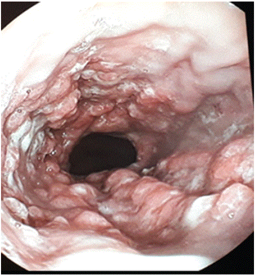
Figure 1: Upper endoscopy: innumerable polypoid and fungating wart-like lesions involving more than 75% of the distal esophagus circumference.

Sabbah Meriam1* Briki Ines1 Bellil Nawel1 Jouini Raja2 Bibani Norsaf1
1Department of gastroenterology, Habib Thameur Hospital, Tunisia*Corresponding author: Sabbah Meriam, Department of gastroenterology, Habib Thameur Hospital, Tunisia, E-mail: sabbah_meriam@yahoo.fr
Extensive forms of squamous esophagealpapilloma are rare. We report a case of 54-year-old papillomatousoesophageal lesion. Patient was negative for HPV infection. He was put on IPP therapy with plans of endoscopic follow up.
This case illustrates the synergic action of chronic mucosal irritation and HPV infection in the development of this tumor. It also shows there should be further screening for other sites of HPV infection if genital warts or other location is noted.
Extensive forms of squamous esophageal papilloma represent rare endoscopic finding and the management is only reported in few case reports of literature [1-3].We report a rare case of 54-year-old woman with a 6 year history of heartburn and recent onset of vomiting with digestive bleeding who presented a papillomatousoesophageal lesion confirmed histologically
A 54-year-old woman presented to the Gastroenterology department for heartburn and two episodes of hematemeses. She reported a 6-year history of heartburn and esophageal dysphagia to solids. Her symptoms worsened recently with the onset of recurrent vomiting associated with epigastric pain. She denied any associated weight loss, changes in her bowel pattern, melena or history of indigestion. Although she showed no symptom of anemia and dizziness, laboratory investigations found a normocytic normochromic anemia with a hemoglobin level of 11g/dl.
Upper endoscopy showed innumerable polypoid and fungating wart-like lesions involving more than 75% of the distal esophagus circumference as well as two longitudinal mucosal erosions greater than five 5mm located between the latter lesions , a small hiatal hernia, as well as a nodular appearance of the duodenal mucosa (Figure 1).
Figure 1: Upper endoscopy: innumerable polypoid and fungating wart-like lesions involving more than 75% of the distal esophagus circumference.
Multiple biopsy specimens were taken from the esophageal lesions as well as a few from the duodenum mucosa and sent for pathological examination.
The biopsy specimens of the sampled esophageal lesions showed squamous papillomas characterized by finger-like projections of a fibrovascular core covered by mature squamous epithelium with no dysplastic cells detected (Figure 2).The duodenal specimens showed an unspecific duodenitis.
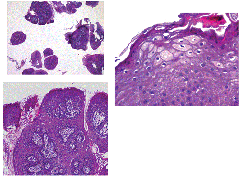
Figure 2: Oeophageal biopsy: squamous papillomas characterized by finger-like projections of a fibrovascular core covered by mature squamous epithelium with no dysplastic cells detected.
Upon further questioning, the patient reported genital warts that have been removed by her dermatologist years ago but the lesions have reemerged. She was also being regularly tested by a pap smear for cervical cancer; the last test is reportedly negative.
The biopsy samples were tested negative for HPV infection using in situ hybridization (ISH)/ p16 immunochemistry expression. We put the patient on proton pump inhibitor (PPI) therapy with plans for endoscopic follow-up.
Esophageal squamous papilloma (ESP) is a rare benign epithelial tumor with a reported prevalence of 0, 01 % -0 ,45% [1].
Typically, these lesions present as a solitary sessile formation with warty-like surface, small in size and mostly in the distal esophagus[2]. Extensive lesions or esophageal papillomatosis (OP) are an even more rare endoscopic finding with only a few case reports described in the literature [3].
ESP can be confused endoscopically with glycogenic acanthosis, verrucouscarcinoma, squamous cell carcinoma. The diagnosis requires biopsies. Although generally asymptomatic, extensive lesions of ESP were associated with dysphagia and peptic disease symptoms such as our case study [1].
The underlying pathogenesis is still controversial but mainly two risk factors have been reported: HPV infection most commonly type 16 but the majority of ESP lesions tested negative [2,4,5] and chronic mucosal irritation associated with reflux or oesophagitis as many cases have shown [6]. Hence, many reports suggest the synergic action might be necessary [2]. In our case, the patient has multiple distal papillomas with a hiatal hernia and mucosal breaks suggesting reflux oeophagitis and biopsy specimens tested for HPV infection were negative.
There is no association between ESP and esophageal cancer [3] although squamous papilloma is considered a premalignant lesion in other mucosal locations (oral, pharyngeal and anogenital. Malignant transformation has been reported however in a few case reports of OP [7,8]. Among the risk factors for malignancy: esophageal stricture, dysphagia, and virulent HPV strains [9].
The management of ESP is not consensual yet in the literature .Small solitary lesions have been successfully treated with endoscopic resection using biopsy forceps, snare polypectomy and cautery and recently radiofrequency ablation [1,10]. During follow up after endoscopy removal examination, one study found recurrenceis uncommon [1]. However, extensive esophageal papillomatosis is still a matter of research due to the paucity of reported cases: several treatment options were suggested mainly protons pumps inhibitors treatment [9], if failed, the endoscopic removal of the largest lesions often requiring endoscopic mucosal resection [11]. Surgical resection has even been advocated when malignancy is suspected [7] or for extensive papillomatosis [12] .If no specific treatment is undertaken, surveillance endoscopy should always be considered given the malignancy potential. However, no specific surveillance strategy has been defined. In our case, the patient is scheduled for endoscopic follow up.
Author’s contribution: Sabbah Meriam, Briki Ines, and Bellil Nawel wrote the manuscript, Jouini Raja Gives the pathology pictures, Bibani Norsaf held the case and performed upper endoscopy
Download Provisional PDF Here
Article Type: CASE REPORT
Citation: Meriam S, Ines B, Nawel B, Raja N, Norsaf B (2020) Oesophagealpapillomatosis in a Patient with History of Genital Warts. J Clin Case Stu 5(4): dx.doi.org/10.16966/2471-4925.209
Copyright: © 2020 Meriam S, et al. This is an open-access article distributed under the terms of the Creative Commons Attribution License, which permits unrestricted use, distribution, and reproduction in any medium, provided the original author and source are credited.
Publication history:
All Sci Forschen Journals are Open Access