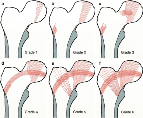
Figure 1: Singh Index grade 1-6 (Reproduced courtesy of Singh M, et al. [20] JBJS Am 1970).


Ong-art Phruetthiphat* Teerapat Tutaworn Yanin Plumarom Thipachart Punyaratabandhu Chaisiri Chaichankul Warat Tassanawipas
Department of Orthopedics, Phramongkutklao Hospital, Bangkok, Thailand*Corresponding author: Ong-art Phruetthiphat, Department of Orthopedics, Phramongkutklao Hospital, Bangkok, Thailand, Tel: (66)81- 4085470; E-mail: ongart-phr1@hotmail.com
Introduction: The incidence of hip fracture in elderly patients has projected to increase world-wide and they were associated with morbidity and mortality. The AAOS 2014 suggested cephalomedullary nail for unstable type of intertrochanteric fracture with a moderate strength of recommendation. However, there was some mechanical failure from Proximal Femoral Nail Anti-rotation (PFNA) blade cut-out in clinical practice after osteoporotic hip fracture fixation. Current literatures lack data demonstrating PFNA with cement augmented blade in clinical study. Therefore, purpose of our study was to undergo the preliminary study whether PFNA with cement augmented blade reduces the prevalence of mechanical failure for intertrochanteric fracture in elderly patients.
Materials and methods: After Institutional Research Board Approval, 10 elderly patients with unstable intertrochanteric fractures and Singh index less than grade 4 underwent PFNA with cement augmented blade was done. The primary outcome was measured by the prevalence of PFNA blade cutout. The secondary outcome was time to union, mortality, and functional outcome (HHS). Cement augmented PFNA group was further compared to the standard PFNA (without cement augmentation) in demographic, comorbidities, mechanical failure, mortality, and one year functional outcome.
Results: All patients underwent cement augmented PFNA fixation and had no mechanical failure. Additionally, there was comparable rate of mechanical failure between groups (0% vs 3.6%, p=1.000). There was similar time to union (9 vs 9 weeks, p=0.517), mortality rate (10% vs 10.7%, p=1.000) and one-year functional outcome (75.0 vs 75.7, p=0.834) between groups.
Conclusion: PFNA with cement augmented blade is safe and this novel technique may be an alternative option for management of unstable type intertrochanteric fracture in elderly patients with discontinuity of principal tensile trabeculae.
Cement augmentation; PFNA; Unstable; Intertrochanteric fracture; Mechanical failure
The incidence of hip fracture in elderly patients has projected to increase worldwide [1-2] and they were associated with morbidity and mortality [3-6]. There were comparable results between intramedullary device and extramedullary device for stable type intertrochanteric fracture [7-10] while intramedullary nail was appropriately applied for unstable intertrochanteric fracture [11,12]. Additionally, the AAOS 2014 suggested cephalomedullary nail for unstable type of intertrochanteric fracture with a moderate strength of recommendation [13] and Socci AR, et al. advocate fixation of 31A2 and 31A3 for unstable type intertrochanteric fracture with intramedullary device [14,15]. The reasons why several studies suggested a cephalomedullary device because they have determined improved walking ability [16] and tend to offer a shorter recovery [17]. However, there was some mechanical failure from Proximal Femoral Nail Anti-rotation (PFNA) blade cut-out in clinical practice after osteoporotic hip fracture fixation because of discontinuity of principal tensile trabeculae (lack of bone trabeculae to prevent blade cut-out). A few literatures have identified potential of Polymethylmethacrylate (PMMA) cement-augmented helical PFNA blades to improve implant stability only in human cadaveric study [18,19]. Moreover, they lack data demonstrating PFNA with cement augmented blade in clinical study. Therefore, purpose of our study was to undergo the preliminary study whether PFNA with cement augmented blade reduces the prevalence of mechanical failure for intertrochanteric fracture in elderly patients.
After obtaining Institutional Research Board Approval (IRB ID S061h/60), 10 elderly patients with unstable intertrochanteric fractures and Singh index [20] less than grade 4 (grade 1-3) underwent PFNA with cement augmented blade were done at orthopedic department, Phramongkutklao hospital, Thailand. Singh index was used for assessment of bone density of femoral head and neck which was identified by Singh index. Singh index of 4 to 6 are normal principal tensile trabeculae while Singh index 1 to 3 are no principal tensile trabeculae, almost loss of principal tensile trabeculae, and discontinuity of principal tensile trabeculae, respectively.
The primary outcome was measured by the prevalence of PFNA blade cut-out or cut-through. The secondary outcome was the functional outcome (HHS) and complications including Venous Thromboembolism (VTE). Functional outcome was categorized into 4 levels: excellent (90-100), good (80-89), fair (70-79), and poor outcome (<70).
A preliminary study of intertrochanteric fractures in elderly patients with low energy trauma, treated with PFNA fixation and cement augmented blade between January 2016 and December 2017 at a single institution were prospectively identified. Hip fracture patients were excluded when they sustained high energy trauma, poly trauma, pathologic fracture, and ballistic injury. Patients who followed up less than 1 year were excluded from the study. Ten cases of low energy intertrochanteric fractures with discontinuity of principal tensile trabeculae (Singh index less than grade 4 as shown in figure 1) underwent PFNA fixation with cement augmentation were done. All patients were collected for demographic data and comorbidities including American Society Anesthesiologist (ASA) classification. Fracture patterns based on Modified AO/OTA 2018 classification were collected in all cases. “Stable type” defined as Modified AO/OTA Type 31 A1.1, A1.2, A1.3. The remaining types (Modified AO/OTA Type 31 A2.2, A2.3, A3.1, A3.2, and A3.3) were “unstable type” as shown in figure 2. All patients were identified for clinical and radiological outcome at 3, 6, and 12 months consequently. We measured quality of reduction (neck-shaft angle, displacement between cortices of proximal and distal fragments in AP and lateral view), Tip and Apex Distance (TAD) and any mechanical failure.

Figure 1: Singh Index grade 1-6 (Reproduced courtesy of Singh M, et al. [20] JBJS Am 1970).
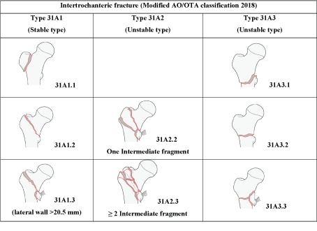
Figure 2: Intertrochanteric fracture (stable and unstable types) by Modified AO/OTA classification 2018.
A case series involving 84 cases of intertrochanteric fracture patients during 2015-2017 underwent fixation with standard PFNA (PFNA without cement augmentation).
Fractures fixation was done by titanium PFNATM nail (DePuy Synthes, Switzerland). All cases were applied on the fracture table in supine position. Closed reduction was done under fluoroscopy. Skin incision was done at lateral aspect of affected hip, guide wire was inserted into the tip of greater trochanter, proximal reaming by hand ream was done, diameter of nail was measured under fluoroscopy and proximal femoral nail was applied into medullary canal while guide wire was removed. Before the application of the helical blade into femoral head, we inserted guide wire into femoral head measured the exact position and length of helical blade in both Anteroposterior (AP) and Lateral (Lat) views. Lateral wall was opened carefully by reaming instrument before blade insertion. We further applied helical blade into the femoral head and it was tightened in the final step. Potential leakage was checked by using a contrast fluid and appropriate syringe (6 milliliters) with Luer lock as shown in figure 3. Contrast fluid approximately 4 milliliters was injected and it was measured under image intensifier to ensure that it was not any leakage into the hip joint. If there is was contrast leakage into the hip joint, the cement augmentation should not be used. The TRAUMACEM™ V+cement kit (DePuy Synthes, Switzerland) is mixed as suggested by manufacture guide and prefilled in needle kit (4 milliliters). The syringe was then attached to side-opening cannula and the cement was injected under fluoroscopic control. Cement augmentation was injected into voiding area of femoral head (at least 2 milliliters), particularly in upper-lateral area of femoral head as shown in figure 4. After finishing injection, the cannula was then removed. The setting time of cement was about 10-15 minutes.

Figure 3: Picture PFNA with cement augmentation (Reproduced courtesy of instrument and implants approved by AO Foundation).
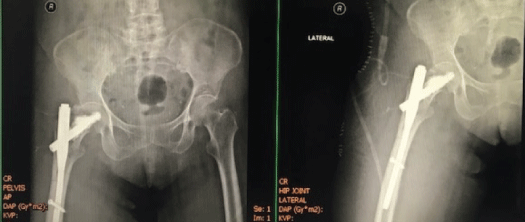
Figure 4: Case example of unstable type intertrochanteric fracture underwent PFNA Fixation and cement augmentation.
Risk of guide wire perforation is very low because we always checked the position of guide wire meticulously by intraoperative fluoroscopy to make sure that guide wire is not perforated. In our case series, we had no guide wire perforation. However, patients can convert into standard PFNA (PFNA without cement augmentation) when guide wire perforates into hip joint.
Patient sustained an unstable type of intertrochanteric fracture was evaluated including preoperative planning by preoperative hip X ray in AP and lateral view (As shown in figure 5).
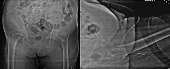
Figure 5: Preoperative AP and lateral radiographs.
Proper analgesic management was done in all patients and they were allowed to bearing weight as tolerate. Deep vein thrombosis prophylaxis at least mechanical pumping was applied during hospital admission. Additionally, patients were allowed to walk with gait aid as soon as possible.
Clinical X rays underwent cement augmented PFNA were followup in clinic immediately, 3 months, and one year as shown in figure 6-8, respectively.
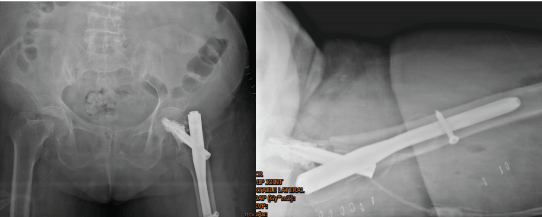
Figure 6: Immediate postoperative AP and lateral radiographs.
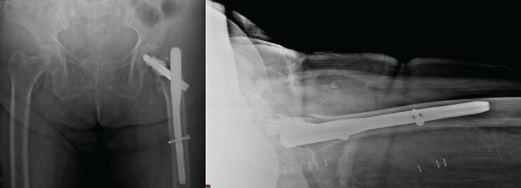
Figure 7: Postoperative AP and lateral radiographs at 3 months follow-up.
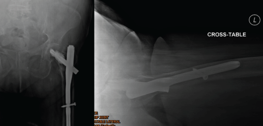
Figure 8: Postoperative AP and lateral radiographs at 1 year follow-up.
All patients were followed up in clinic at 2 weeks, 6 weeks, 3 months, 6 months, and 1 year. Radiographic measurements were done by two orthopedic traumatologists. Antero-posterior (AP) and lateral radiographs (Lat) assessed by PACS software were used for quality of fracture reduction, Neck Shaft Angle (NSA), Tip Apex Distance (TAD) and any implant failure (PFNA blade cut-out was defined as perforation of the helical blade through the superior cortex of the femoral head or femoral neck; PFNA blade cut-through was defined as helical blade migrates centrally into hip joint). The primary outcome was measured by the prevalence of PFNA blade cut-out or cut-through. The secondary outcome was time to union (weeks), functional outcome (Harris Hip Score- HHS), any medical complications including Venous Thromboembolism (VTE), surgical complication, and mortality.
Time to union was classified into 2 types: clinical union and radiographic union. Clinical union was defined by clinical and radiographic measurement: patient can partially bear weight without pain and the radiograph demonstrates incomplete obliteration of the fracture line. Radiographic union was defined by complete obliteration of the fracture line on the radiograph [21].
Harris Hip score (HHS) is composed of many aspects: Pain (44 points), limp (11 points), support (11 points), distance walked (11 points), sitting (5 points), enter public transportation (1 point), stair (4 points), put on shoes and socks (4 points), absence of deformity (4 points), and range of motion (5 points). Zero point’s means the lowest hip score while one hundred points means the maximal hip score. HHS was measured into two aspects in all patients: pre-fracture state by interview and postoperative state at one year follow-up by examination in clinic. Functional outcome (HHS) was categorized into 4 levels: excellent (90-100), good (80-89), fair (70-79) and poor outcome (<70).
Surgical complications (mechanical failure including PFNA blade cut-out, blade cut through, and varus collapse) were recorded.
We further compared the demographic data and comorbidities between cement augmented PFNA (n=10) and standard PFNA (without cement augmentation) (n=84). Additionally, time to union, complications (mechanical failure including mortality), and one year functional outcome assessed by HHS were compared between groups.
Demographic data was recorded for each patient and analyzed using STATA Version 12. Data were summarized using descriptive statistics (number of patients and mean ± SD). Postoperative fracture displacement (Gap and Step), Neck Shaft Angle, and Tip Apex Distance during admission (gap and step in AP and lateral views) (millimeters) were shown in mean ± SD. Preoperative and postoperative functional outcome (HHS) at one year were compared by Paired T-test. Age and Harris Hip Score were assessed by Independent t-test while Fisher’s exact test was used for gender, ASA class, modified AO/ OTA classification, mechanical failure, and mortality. CCI (Charlson Comorbidity Index) and time to union were evaluated by MannWhitney U-test. P-value less than 0.05 was considered a significant difference.
The average age was 82.5 years old (71-94). Female was predominantly in our case series. Underlying diseases and ASA (American Society of Anesthesiologists) classification of all patients were shown in table 1. Almost all patients had high Charlson Comorbidity Index (7 out of 10 had CCI>4). Comparison of demographic data and comorbidities between cement augmented PFNA and standard PFNA were shown in table 2. There was no difference in demographic data and comorbidities between groups.
| Cases | Status | Age (years) | Gender | Site | Underlying disease | ASA classification | Charlson Comorbidity Index |
| 1 | Alive | 78 | Female | Left | HT | 2 | 4 |
| 2 | Alive | 84 | Female | Left | DM2/HT/DLP/ Hyperthyroidism | 3 | 6 |
| 3 | Alive | 86 | Female | Right | HT/DLP/IHD | 3 | 6 |
| 4 | Alive | 71 | Female | Right | DM2/HT/Old CVA | 3 | 8 |
| 5 | Alive | 77 | Female | Right | HT/DLP | 2 | 4 |
| 6 | Alive | 72 | Female | Right | HT | 2 | 4 |
| 7 | Alive | 82 | Female | Left | DM2/HT/DLP/Old CVA | 3 | 8 |
| 8 | Decease* | 92 | Male | Right | BPH | 2 | 5 |
| 9 | Alive | 94 | Male | Left | HT/DLP | 2 | 6 |
| 10 | Alive | 89 | Male | Right | DM2/HT/DLP/ TVD/CKD | 3 | 8 |
| *Death caused by pneumonia/sepsis; HT=Hypertension; DM2=Type II Diabetes Mellitus; DLP=Dyslipidemia; CVA=Cerebrovascular Accident; BPH=Benign Prostatic Hypertrophy; CKD=Chronic Kidney Disease; ASA=American Society of Anesthesiologists |
|||||||
Table 1: Patient’s demographic data and comorbidities of unstable intertrochanteric fracture underwent PFNA with cement augmentation.
| Demographic data | Cement Augmented PFNA (n=10) | Standard PFNA (n=84) | P-value |
| Gender | 1.000 | ||
| Male | 2 (20.00) | 18 (21.43) | |
| Female | 8 (80.00) | 66 (78.57) | |
| Age (years) (Mean ± SD) | 82.50 ± 7.98 | 81.64 ± 7.59 | 0.738 |
| ASA class | 0.698 | ||
| I | 0 (0%) | 9 (10.71) | |
| II | 5 (50%) | 44 (52.38) | |
| III | 5 (50%) | 31 (36.90) | |
| CCI (Mean)(Min-Max) | 5.5 (3-8) | 4.5 (1-9) | 0.198 |
| ASA class=American Society of Anesthesiologist Classification; CCI=Charlson Comorbidity Index | |||
Table 2: Comparison of demographic data and comorbidities between cement augmented PFNA and standard PFNA.
All patients treated with cement augmented PFNA fixation were fallen into unstable type of intertrochanteric fracture. Patients with Modified AO/OTA 31-A2.2 were 80% while the others (20%) were Modified AO/OTA 31-A2.3 (10%) and 31-A3.2 (10%). The comparison of fracture patterns by Modified AO/OTA 2018 between cement augmented PFNA and standard PFNA were shown in table 3.
| Fracture patterns | Cement Augmented PFNA (n)(%) | Standard PFNA (n)(%) | P-value |
| A1.1 | 0 (0%) | 0 (0%) | NA |
| A1.2 | 0 (0%) | 8 (9.5%) | 0.593 |
| A1.3 | 0 (0%) | 9 (10.7%) | 0.590 |
| A2.2 | 8 (80%) | 41 (48.8%) | 0.094 |
| A2.3 | 1 (10%) | 12 (14.3%) | 1.000 |
| A3.1 | 0 (0%) | 1 (1.2%) | 1.000 |
| A3.2 | 1 (10%) | 3 (3.6%) | 0.367 |
| A3.3 | 0 (0%) | 10 (11.9%) | 0.593 |
Table 3: Comparison of fracture patterns between cement augmented PFNA and standard PFNA by modified AO/OTA 2017 classification.
According to the cement augmented PFNA group, five patients (50%) were perfect reduction (no gap and step in both AP and lateral views) while the others (50%) were reduced with minimal displacement (gap/step less than 5 millimeters). Six cases (60%) were reduced within normal neck shaft angle (NSA) while remaining cases (40%) were fallen into coxa valga. (Table 4).
| Cases | Displacement (millimeters) AP view | Displacement (millimeters) Lateral view | Neck Shaft Angle (Degree) | Tip Apex Distance (millimeters) | ||||||
| Gap | Step | Gap | Step | AP view | Lateral | Summation | ||||
| 1 | 0 | 5 | 0 | 5 | 132.0 | 10.7 | 11.7 | 22.4 | ||
| 2 | 0 | 0 | 0 | 0 | 129.3 | 13.4 | 12.8 | 26.2 | ||
| 3 | 0 | 0 | 0 | 0 | 142.6 | 7.3 | 8.3 | 15.6 | ||
| 4 | 5.6 | 0 | 3.8 | 0 | 141.2 | 6.5 | 9.1 | 15.6 | ||
| 5 | 0 | 0 | 0 | 0 | 130.5 | 14.2 | 11.3 | 25.5 | ||
| 6 | 0 | 0 | 0 | 0 | 132.1 | 7.4 | 8.1 | 15.5 | ||
| 7 | 0 | 0 | 3.3 | 4.8 | 130.2 | 14.7 | 13.2 | 27.9 | ||
| 8 | 0 | 3.9 | 1.9 | 0 | 137.7 | 12.4 | 11.6 | 24.0 | ||
| 9 | 0 | 0 | 0 | 2 | 135.6 | 8.4 | 10.6 | 19.0 | ||
| 10 | 1.2 | 1.0 | 1.0 | 1.3 | 134.2 | 10.6 | 10.7 | 21.3 | ||
| Mean | 0.68 | 0.99 | 1 | 1.31 | 134.54 | 10.56 | 10.74 | 21.3 | ||
| SD | 1.77 | 1.87 | 1.49 | 2.02 | 4.66 | 3.05 | 1.76 | 4.69 | ||
| Coxa vara was defined as Neck Shaft Angle (NSA) <120 degree (°); Normal NSA was defined as NSA between 120-135° while Coxa valga was defined as NSA >135° | ||||||||||
Table 4: Postoperative fracture displacement (Gap and Step), neck shaft angle, and tip apex distance during admission.
Mean Tip Apex Distance (TAD) ± Standard deviation (SD) in AP view, lateral view, and summation of TAD were 10.56 ± 3.05 millimeters (mm), 10.74 ± 1.76 mm, and 21.30 ± 4.69 mm, respectively (Table 4).
Comparison demographic data and comorbidities between cement augmented PFNA and standard PFNA were shown in table 2. There was no difference in gender, age, ASA classification, and CCI.
Clinical and radiographic time to union was comparable between groups as shown in table 5.
| Parameters | Cement Augmented PFNA (n=10) | Standard PFNA (n=84) | P-value |
| Time to union (weeks)* | |||
| Clinical union | 6 (4-8) | 6 (2-9) | 0.406 |
| Radiographic union | 9 (6-18) | 9 (6-19) | 0.517 |
| Complication | |||
| Mechanical Failure | 0 (0%) | 3 (3.6%) | 1.000 |
| Mortality | 1 (10%) | 9 (10.7%) | 1.000 |
| Harris Hip Score | |||
| Pre-injury | 79.33 ± 7.28 | 68.37 ± 15.45 | 0.019 |
| Postoperative at one year | 75.00 ± 5.44 | 75.67 ± 11.43 | 0.834 |
| *=Median (Minimum-Maximum) | |||
Table 5: Comparison of time to union, complications and functional outcome between cement augmented PFNA and standard PFNA.
Pre-injury status in all patients were partially dependent with assistive device (20% with 1-point cane, 30% with 3-points cane, and 50% with walker) while postoperative activity in all patients after surgical fixation at one year follow-up was partially dependent with walker. Unfortunately, 1 patient was died from medical complication at 7 months after surgery with pneumonia and sepsis, as shown in table 6.
| Cases | Activity level | Functional Outcome (HHS) at one year | Complications | |||
| Preoperative | Postoperative | Preoperative | Postoperative | Medical* | Surgical** | |
| 1 | 3 points cane | Walker | 77 | 72 | No | No |
| 2 | 3 points cane | Walker | 78 | 72 | No | No |
| 3 | 1 point cane | Walker | 83 | 78 | No | No |
| 4 | Walker | Walker | 69 | 67 | No | No |
| 5 | Walker | Walker | 70 | 70 | No | No |
| 6 | 1 point cane | Walker | 91 | 83 | No | No |
| 7 | Walker | Walker | 77 | 74 | No | No |
| 8 | Walker | Died | 78 | Died | Yes | No |
| 9 | Walker | Walker | 78 | 76 | No | No |
| 10 | 3 points cane | Walker | 75 | 70 | No | No |
| Mean ± SD | 77.55 ± 6.59*** | 73.55 ± 4.85*** | ||||
| HHS: Excellent (90-100), Good (80-89), Fair (70-79), and Poor (<70) *No patients have short term medical complication including venous thromboembolism within 3 months. However, one patient was died at 7 months after surgery because of pneumonia. **No patients have surgical complication including surgical site infection and mechanical failure (Cut-out, Cut-through blade, or varus collapse) ***Preoperative and postoperative functional outcome (HHS) at one year was compared by Paired T-test. P-value was 0.001 (Statistical significant) |
||||||
Table 6: Patient’s outcome assessed by Harris Hip Score (HHS) and complications following PFNA with cement augmentation.
Previous our case series (Standard PFNA for intertrochanteric fracture in elderly patients during 2015 to 2017) had a small number of mechanical failure (3 out of 84 cases; 3.6%) (Table 5). Two PFNA blade cut-out and one PFNA blade cut-through. All of failure cases were later converted into bipolar hemiarthroplasty. On the contrary, none of all patients in cement augmented PFNA group were assessed as having mechanical failure caused by PFNA blade cut-out/cutthrough and having venous thromboembolism in our case series as shown in table 7.
| Grade | Number of Patients (%) | |
| Functional outcome (Harris Hip score at one year) | Excellent (90-100) | 0 (0%) |
| Good (80-89) | 1 (11.11%) | |
| Fair (70-79) | 7 (77.78%) | |
| Poor (<70) | 1 (11.11%) | |
| Complications | Medical | 1 (10%)* |
| Surgical (Mechanical failure) | 0 (0%) | |
| *Rate of Medical complication was 10% at one year after surgery (one patient was died because of pneumonia/sepsis at 7 months follow-up) | ||
Table 7: Functional outcome and complications after PFNA Fixation with cement augmentation in unstable type intertrochanteric fracture.
Almost all patients (88.89%) had significantly lower functional score after postoperative at one year assessed by HHS comparing to pre-injury status as shown in table 6. Average preoperative and postoperative HHS were 77.55 ± 6.59 and 73.55 ± 4.85, respectively, and there was significantly different (p-value 0.001).
There was one good functional outcome (11.11%), 7 fair functional outcome (77.78%), and one poor functional outcome (11.11%) at one year follow-up while they had one excellent (10%), one good (10%), seven fair (70%), and one poor (10%) preoperative functional outcome (Table 6). One year functional outcome assessed by HHS (75.0 vs 75.7, p=0.834) was comparable between groups (Table 5).
Hip fracture in Elderly was associated with increased morbidity and mortality, also cost of treatment [3-6]. Operative treatment is generally a standard treatment because it provided earlier ambulation, faster recovery, and lower complications. As we know, intramedullary nail was appropriately applied for unstable intertrochanteric fracture [11,12], however, failure rate requiring reoperation was reported up to 5.7% [22]. Our case series performed standard PFNA fixation (without cement augmentation) between 2015 until 2017 for intertrochanteric fracture in elderly patients (n=84). Even though almost of all patients had successful outcome, 3.6% (3 out of 84 cases) had mechanical failure (2 PFNA blade cut-out and 1 PFNA blade cut-through). All of failure cases were later converted into bipolar hemiarthroplasty. This is the reason why we performed the preliminary study of PFNA with cement augmented blade for unstable type of intertrochanteric fracture in elderly patients.
Current literature has demonstrated that cement augmented PFNA provided short-term functional result [23] and cement augmentation gives construct much more stability because of larger bone-implant interface [24-26]. However, our study was comparable rate of mechanical failure (0% and 3.6%, p=1.000) between cement augmented PFNA and standard PFNA groups.
An average Charlson Comorbidity Index in our series was much higher than those population defined by multicenter randomized controlled trial of PFNA with cement augmentation (5.9 vs 2.0) even though mean age of population in our study was slightly lower than the Kammerlander C, et al. [27] Study (82.5 vs 86.1 years old). This is the reason why an ambulatory status postoperatively in our series was significantly poorer than previous study.
The primary outcome in this study was to identify the prevalence of PFNA blade cut-out and cut-through. We observed no any mechanical failure (0%) including PFNA blade cut-out and cut-through. Several reasons why this study gained a successful outcome in all cases because 1) an average TAD in our study was 21.3 mm which was well within an acceptable range (20-30 mm) as defined in previous studies [28], 2) an appropriate NSA [60% normal NSA and 40% coxa valga (stable pattern of fracture reduction)], 3) Good quality of reduction (50% had no displacement and 50% had minimal displacement), and 4) additional cement augmentation completely filled in discontinuity area of principal tensile trabeculae.
The secondary outcome including functional outcome was assessed by HHS, complication and mortality. Even though 44.44% percent of our patients reached preoperative functional status at 1 year followup (ambulate with walker), most patients in this study (8 out of 9) (88.89%) had lower functional outcome (assessed by HHS) than preinjury status with statistically difference. However, walking ability in our study was comparable with previous studies demonstrating a high rate of dependence (80% were using a walking aids at 1 year follow-up, 16% were institutionalized, and 11% were bed-ridden postoperatively) [29-31].
Another concern was complication and mortality, Hisatome T, et al. [32] report influence of exothermic reaction from cement when placed in subchondral bone. Likewise, Fliri L, et al. [33] demonstrated that TRAUMACEM V+TM can provide up to 41°C at cement-bone interface when using a volume of 3 millimeters around the blade. Nonetheless, there is no complication related to cement augmentation in our study such as osteonecrosis of femoral head, similarly to another study [27,34]. One year mortality rate in our study was only 10% that was lower when compare to recent systematic review (22%) [35] and the Asian population (17.9%). This might be caused by small sample in our series.
Our preliminary study has some strength: Firstly, we included only unstable type of intertrochanteric fracture classified by Modified AO/ OTA 2018. Secondly, our study identified the clinical results with a prospective design. However, this study had a limitation: this study had small number of population because of preliminary reported. Besides, more populations were needed to actually identify indication for cement augmentation and further randomized studies were needed.
Both cement augmented PFNA and standard PFNA have gained successful outcome with similarly small numbers of mechanical failure. Interestingly, PFNA with cement augmented blade is safe and this novel technique may be an alternative option for management of unstable type intertrochanteric fracture in elderly patients with discontinuity of principal tensile trabeculae.
Download Provisional PDF Here
Article Type: RESEARCH ARTICLE
Citation: Phruetthiphat OA, Tutaworn T, Plumarom Y, Punyaratabandhu T, Chaichankul C, et al. (2019) Proximal Femoral Nail Anti-rotation with Cement Augmentation Reduces the Prevalence of Mechanical Failure for Unstable Type Intertrochanteric fracture in Elderly Patient: A Preliminary Study. J Clin Case Stu 4(3): dx.doi.org/10.16966/2471-4925.192
Copyright: © 2019 Phruetthiphat OA, et al. This is an open-access article distributed under the terms of the Creative Commons Attribution License, which permits unrestricted use, distribution, and reproduction in any medium, provided the original author and source are credited.
Publication history:
All Sci Forschen Journals are Open Access