Figure 1: X-Ray: lung mass on the upper left lobe


Victoria Juarez San Juan* Pedro Rodríguez Suárez Agar Santana Jorge Freixinet Gilart
Almansa 11 4º P6, Las Palmas de Gran Canaria, Spain*Corresponding author: Victoria Juarez San Juan, Thoracic surgery resident (third year), Almansa n 11 4° p6 Cp: 35010 las Palmas de gran canaria, Spain, Tel: +34635454342; E-mail: vito.juarezsanjuan@gmail.com
Osteoblastoma is a rare primary benign bone tumor. The peak incidence of this neoplasm is in the first three decades of life. Men are more susceptible than women, with a ratio of 2,5:1[1].
It generally occurs in the axial skeleton, affecting mainly posterior elements of the vertebra. We report a case of a 26-year-old man with a benign osteoblastoma in a rare location.
Osteblastoma is a benign solitary bone tumor characterized by the formation of osteoid and primitive woven bone. It is extremely rare, representing less than 1% of primary bone tumors [2]. It may be locally aggressive. Usually occurs in young men between the second and third decade of life [3] and it is based predominantly in the axial skeleton and long bones. The most striking symptoms are chest pain and neurological disorders. We report a case of a 26-year-old man with a benign osteoblastoma in a rare location.
A 26 years-old-man suffering from persistent and disbling chest pain, loss of strength and tenderness in the left upper limb, accompanied by ipsilateral ptosis was examined.
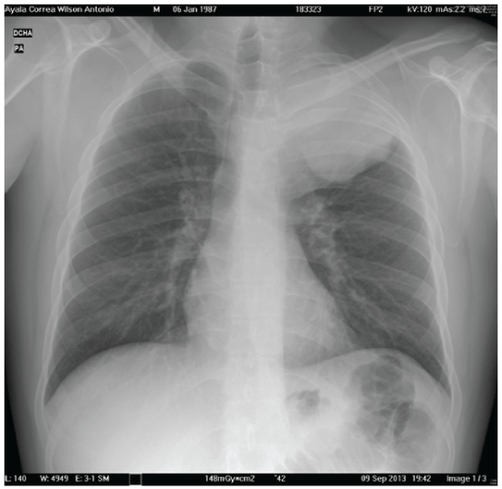
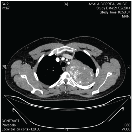
Analytical studies including blood count and biochemistry were normal. The plain radiography and CT scan showed the presence of a lung mass on the upper left lobe of 10 × 12 × 10 cm, with extensive contact to the upper mediastinum and extending into the spinal canal, destroying the mediolateral portion of the vertebral body, transverse process of D3 and posterior portion of the third left rib, showing no pathologic lymph nodes (Figures 1 and 2). Preoperative mass biopsy was done guided by ultrasound and it showed histological and immunohistological findings compatible with giant cell tumor. Surgical resection of the mass was initially discarded, opting for chemotherapy (denosumab) for about a year, with monitoring and radiological control. After lack of radiological tumor response, the patient was sent to our department to assess the surgical resectability of the mass.

Figure 1: X-Ray: lung mass on the upper left lobe

Figure 2: CT scan: lung mass on the upper left lobe involving the spinal canal and destroying the D3 vertebral body and posterior portion of the third left rib
The study was completed with a PET-CT that showed a mass uptake of the maximum SUV of 9.7 and showing infiltration of the ipsilateral 3rd vertebra (maximum SUV of 6.9) and the 3rd left rib (maximum SUV 6.3). MRI also revealed relevant findings such as the contact with the mediastinum and the descending aorta over 3 cm, and an intimate contact with the subclavian vessels, losing plane along 1.3 cm. The mass had made contact with costotransverse articulation joint D3 and introduced into the D3 – D4 hole.
Because of the suspected infiltration of the left subclavian artery, a stent was placed prior to surgery by the vascular surgery team .We decided to make a surgical resection of the mass by a left posterolateral thoracotomy since its the most frequent incision in daily thoracic procedures. The patient was placed in the apropiated complete lateral decubitus position with proper padding to the elbows and knees. The bloc resection of the tumor was performed (3rd and 4th costal arches and complete corpectomy of the 3rd thoracic vertebra) then a mesh clindro plate and a screw were placed.
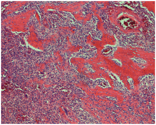
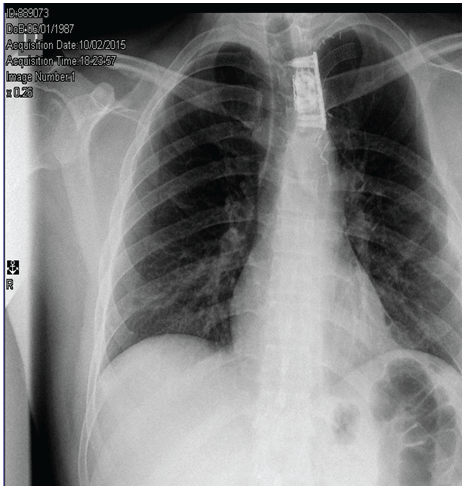
Histological examination showed the presence of large amounts of new osteoid material accompanied by small bone spicules and giant cells (Figure 3). These findings were consistent with a Thoracic Osteoblastoma. Adjuvant therapy was not necessary.After one year of the surgery, the pain has completely dissapeared and the patient is in good general condition, requiring the use of a back brace (Figure 4).

Figure 3: Hystologic Findings: large amounts of new osteiode material accompanied by small bone spicules and giant cells

Figure 4: X-Ray: post surgery
Osteoblastoma is an extremely rare primary bone tumor. It represents 1% of bone tumors and 2-3% of benign primary bone tumors which affect men more frequently. It has a peak incidence between 10 and 30 years. It was first described by Lichtenstein in the mid-twentieth century as osteogenic fibroma of bone. It has been considered a giant osteoid osteoma as both have similar histologic characteristics [4].
It is usually located in the axial skeleton. The most common site is the posterior elements of the vertebrae, rarely affecting vertebral bodies. It generally behaves as a benign tumor but with rapid growth, damaging the bone and infiltrating adjacent structures. Aggressive variations that can metastasize have been described, even having similar behavior to an osteosarcoma.
The most common symptom of this tumor is the pain which does not get worse at night, and its not relieved by aspirin. This is an important difference from osteoid osteoma. Osteoblastoma with a spinal affection can cause scoliosis, neurological symptoms, gait disturbance and breathlessness when there is thoracic involvement.
The radiographic images are eccentric expansive osteolysis surrounded by a zone of variable thickness sclerosis, reaching above 2 cm sizes. MRI should be performed whenever there is suspicion of soft tissue extension or relationship with neurovascular structures [5]. Our patient’s chest- ray revealed typical findings of osteoblastoma
Osteoblastoma can be easily confused with other bone tumor lineage such as osteoid osteoma, osteosarcoma, giant cell tumor, and aneurysmal bone cyst.
Definitive diagnosis was based on the histological findings that reveal mineralized bone spicules, osteoblast-like cells and a large amounts of osteoid and bone .
Treatment includes complete resection of the tumor to prevent recurrence and malignant transformation. Our decision, even without a clear histological diagnosis, was based on the persistence over time of the mass and the need for diagnosis and surgical treatment, which was performed with a single surgery.
I sincerely congratulate the coauthors on the results of their surgical skills in this case.
Download Provisional PDF Here
Article Type: Case Report
Citation: San-Juan VJ, Suárez PR, Santana A, Gilart JF (2017) 26 Years Old Men with Persistent Back Pain: A Thoracic Osteblastoma. J Clin Case Stu 2(3): doi http://dx.doi.org/10.16966/2471-4925.139
Copyright: © 2017 San-Juan VJ, et al. This is an open-access article distributed under the terms of the Creative Commons Attribution License, which permits unrestricted use, distribution, and reproduction in any medium, provided the original author and source are credited.
Publication history:
All Sci Forschen Journals are Open Access