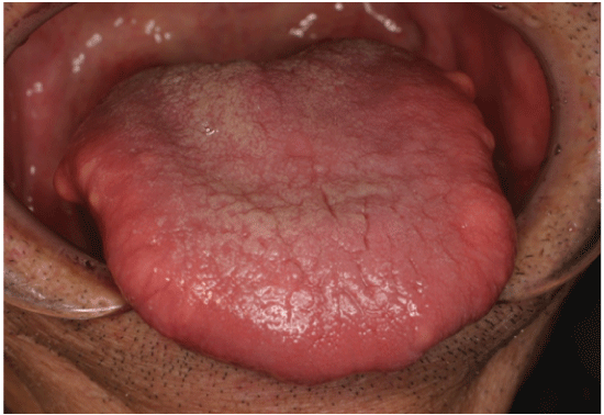
Figure 1: An intraoral view shows many yellowish tumors of the bilateral tongue.


Goro Kawasaki1* Saki Hayashida1 Izumi Yoshitomi2 Syuichi Fujita3 Masahiro Umeda1
1Department of Clinical Oral Oncology, Unit of Translational Medicine, Nagasaki University Graduate School of Biomedical Sciences*Corresponding author: Goro Kawasaki, DDS, PhD, Department of Clinical Oral Oncology, Unit of Translational Medicine, Nagasaki University Graduate School of Biomedical Sciences, 1-7-1 Sakamoto, Nagasaki, 852-8588, Japan, Tel: +81-95-819-7698; Fax: +81-95-819-7700; E-mail: gkawa@nagasaki-u.ac.jp
Symmetric lipomatosis of the tongue is a rare condition. We present a rare case of intramuscular lipomatosis of the tongue in a 70-yearold male. A clinical diagnosis of symmetric lipomas of the tongue was made. The tumors of the bilateral tongue were resected under general anesthesia. The pathological diagnosis was symmetrical lipomatosis of the tongue. There were no signs of recurrence 3 years after the operation.
Lipomatosis; Tongue; Symmetric
Lipomatosis is a condition characterized by benign tissue tumors on many parts of the body. It commonly affects the upper back and shoulder, but also occurs in the oral cavity at lower rates [1]. Multiple tongue lipomas, particularly of a symmetric tumor formation, are called symmetric lipomatosis of the tongue [1-3]. Lipomatosis of the tongue is extremely rare [4] (Figure 1).

Figure 1: An intraoral view shows many yellowish tumors of the bilateral tongue.
Here, a case of symmetric lipomatosis of the tongue with intramuscular invasion is presented, and the current literature is reviewed.
In November 2011, a 70-year-old man was referred to Nagasaki University Hospital complaining of bilateral swelling of the tongue. He had been aware of the swelling for many years, and he finally consulted his dentist.
On clinical examination, no tumor masses could be identified on the trunk, head, neck, or extremities. The tongue was diffusely enlarged and many small tumor-like lesions of yellowish color were observed bilaterally on the tongue. There was no dysphagia, ankyloglossia, or dyspnea. The patient had a history of hypertension, cerebral palsy after head injury, and moderate alcohol consumption for more than 50 years.
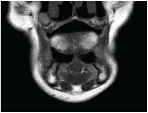
Magnetic resonance imaging (MRI) demonstrated multilocular lesions having high signal intensity on T1W1 bilaterally on the tongue, and the lesion was diminished by fat suppression (Figure 2).

Figure 2: T1W2 MRI shows diffuse high intensity area on the tongue.
Clinical diagnosis was symmetric lipomatosis of the tongue.
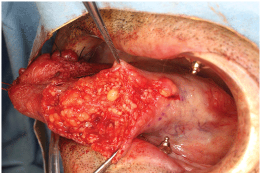
The tumors were resected under general anesthesia. Adipose tissue proliferation was unencapsulated (Figure 3).

Figure 3: Yellowish adipose tissues were seen just beneath the epithelium on the lateral border of the tongue. The adipose tissues infiltrated the lingual muscles.
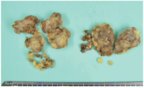
Histopathological findings revealed adipose tissue consisting of mature adipocytes infiltrating between muscle fibers, confirming the diagnosis of symmetrical lipomatosis of the tongue (Figures 4 and 5).

Figure 4: The excised tumors showed no capsule formation.

Figure 5: Pathological findings: Adipose tissues were interspersed with the muscle fibers (H-E staining, × 100).
Lipoma of the tongue is rare and often presents as a single, superficial, pedunculated, and sessile lesion. Presentation as multiple tumors, infiltrating tumors, or macroglossia is infrequent [5]. Diagnosis of lipoma of the tongue is essentially clinical, but radiological investigations, especially magnetic resonance imaging (MRI), may be helpful in diagnosis [5,6].
Lipomas are classified into 5 categories: (1) lipoma, (2) variants of lipoma, (3) heterotopic lipoma, (4) infiltrating/diffuse, neoplastic/nonneoplastic proliferation of mature fat, and (5) hibernoma [7,8]. The fourth group includes benign symmetric lipomatosis, diffuse lipomatosis, and pelvic lipomatosis. Lipomatosis is characterized by multiple sites of involvement, invasiveness, and absence of encapsulation of the adipose tissue [4].
Our case had these characteristics, and thus we made a diagnosis of lipomatosis.
Symmetric lipomatosis of the tongue is reported to be an extremely rare disease [9]. Calvo-Garcia et al. [4] described only 6 cases of symmetric lipomatosis, including one case reported in the English literature. On the other hand, tongue symmetric lipomatosis has been reported more in Asia [10]. We reviewed 28 cases of symmetric lipomatosis of the tongue in the Japanese literature [11-29]. All of the patients were male, and 17 of 28 patients suffered from alcoholic hepatitis and/or alcoholism.
Acetyl CoA, an organic compound, is a metabolite of alcohol in the liver, and it produces neutral lipids [30]. It is generally known that an increase of acetyl CoA is associated with fatty deposition [30]. In our case series, there were many cases of alcoholic hepatitis and alcoholism; hence, we suggest that symmetric lipomatosis of the tongue is associated with alcohol metabolic deposition.
With regard to treatment, the gold standard is complete surgical resection [1]. In our case series, there was surgical resection of the tumor in 18 of 28 cases. The reasons for non-surgical treatment in our case series were poor general condition, unwillingness of the patients to undergo surgical treatment, and lack of subjective symptoms. If a patient has a functional disorder, such as dysarthria and/or dysphagia, surgical treatment is recommended. In the present case, the patient desired surgery because of difficulty in food intake due to tongue enlargement. Since surgery, there have been no signs of recurrence.
There are no conflicts of interest to report.
Download Provisional PDF Here
Article Type: Case Report
Citation: Kawasaki G, Hayashida S, Yoshitomi I, Fujita S, Umeda M (2017) Symmetric lipomatosis of the Tongue: Report of a Case. J Clin Case Stu 2(1): doi http://dx.doi.org/10.16966/2471-4925.138
Copyright: © 2017 Kawasaki G, et al. This is an open-access article distributed under the terms of the Creative Commons Attribution License, which permits unrestricted use, distribution, and reproduction in any medium, provided the original author and source are credited.
Publication history:
All Sci Forschen Journals are Open Access