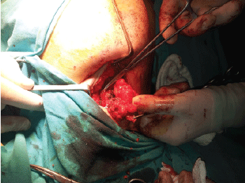
Figure 1: Surgical resection, lesion with skin


Erol Kilic1 Ibrahim Yetim1* Cem Oruc1 Mustafa Ugur1 Hasan Gokce2
1Department of General Surgery, Mustafa Kemal University, Serinyol, Hatay, Turkey*Corresponding author: Ibrahim Yetim, Department of General Surgery, Mustafa Kemal University, Medical School, Serinyol, Hatay, Turkey, Tel: 5325060009; Fax: 00903262291900; E-mail: yetim54@gmail.com
Primary ectopic breast carcinoma (PEBC) is a very rare and progressive case, most commonly encountered in the advanced stage. Most of the cases develop from the ectopic breast tissue (EBT), and it may occur along the milk line within the area from the axilla to the external genital region. EBT is observed in the 6% of the female population and is mostly encountered in the axilla. Clinically, the most frequently encountered type of PEBC is the one which originates from the EBT located in the axillary and it constitutes 70% of all PEBCs. Histopathologically, the most common type is invasive ductal carcinoma (72%). Mammography and ultrasound-guided fine needle aspiration (FNA) employed in the diagnosis of PEBC and also in the differential diagnosis of lipoma, lymphadenitis, metastatic carcinoma and the lesions such as hidradenitis suppurativa. In the surgical treatment of PEBC is recommended the resection of the tumor tissue with the skin together and the dissection of the regional LNs. Loco-regional approach (Ro local excision+axillary dissection+radiotherapy) has been considered to be the most appropriate method in PEBC treatment and has been employed for 15 years.
Primer ectopic breast carcinoma; Ectopic breast tissue; Diagnosis; Treatment
Primary ectopic breast carcinoma (PEBC) is a very rare and progressive case most commonly encountered in the advanced stage. Most of the cases develop from the ectopic/accessory breast tissue (EBT). Embryologically, EBT originates from ectoderm thickening and it may occur along the milk line within the area from the axilla to the external genital region. In this study we aimed to discuss the epidemiology, clinic and treatment of PEMC originating from the EBT in axilla, which is very rare and follows a progressive clinical course.
The 56-year-old female patient reported that she had felt firmness in the right axilla for about 5 years and a skin retraction over the past year. Physical examination revealed an about 3 × 4 cm palpable mass in the right axilla, causing the retraction of the skin. In the physical examination of both breasts; left axilla, bilateral supraclavicular fossa and bilateral servicolateral lymph node areas were normal.
In USG, 23 × 20 mm lesion in right axilla (brads V) determined. Preoperative stage were T2N2M0 (Mammography were normal). Surgical resection included loco-regional surgery (Ro resection+axillary lymphadenectomy) and lesion with skin (Figure 1). Histopathological study revealed an invasive ductal carcinoma (ER+3, PR+3, Her2/neu (c-erbB2) score: 1+, Ki-67 %10, E-Cadherin+). The diameter of the tumor was 1.7 cm. It was Grade 2 according to the MBR (Modified Bloom Richardson) and lymphovascular invasion and perineural invasion were positive. There were 2 metastatic lymph nodes. Surgical margins were tumor negative.

Figure 1: Surgical resection, lesion with skin
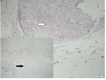
Postoperative, to examination of a possible distant organs and bone metastases implemented FDG-PET. FDG-PET revealed involvement in C5, T10 and L2 vertebrates (degenerative changes) and in the right axilla (surgical changes). Postoperative stages of tumor were T1N1M0. Ipsilateral mastectomy did not implemented, received postoperative radiotherapy and chemotherapy/ hormonotherapy of patient is continued (Figure 2). Before this article is written, informed consent was obtained from the patient.

Figure 2: Nuclear painting with estrogen and progesterone
Ectopic breast tissue is most commonly encountered in the axilla, but can also be seen in the face, chest wall, posterior neck area, back, hips, flank area, vulva, femoral area, shoulders and upper extremities [1]. Clinically, the most frequently encountered type of PEBC is the one which originates from the EBT located in the axillary and it constitutes 70% of all PEBCs [2]. Histopathologically, the most common type is invasive ductal carcinoma (72%). Invasive lobular carcinoma and medullary carcinoma are less common (12%). Other carcinoma types constitute the remaining 16% [3]. In anamnesis, PEBC is defined as a progressive tumor with unilateral subcutaneous location, but having no relation with the local symptoms. Predisposing factors in primary breast carcinoma (PBC) such as familial history or exposure to radiation can also be effective in PEBC [4]. Clinically, partial/complete areola and nipple belonging to EBT can also be observed [4]. There was no nipple or areola in our case. Along with the two breasts and axillaries, bilateral supraclavicular fossa and bilateral laterocervical lymph nodes should also be examined in PEBC patients [5]. In case, carcinoma was originating from EBT in axilla and its histopatological diagnosis was invasive ductal carcinoma. Mammography and ultrasound-guided fine needle aspiration (FNA) employed in the diagnosis of PEBC and also in the differential diagnosis of lipoma, lymphadenitis, metastatic carcinoma and the lesions such as hidradenitis suppurativa [1,6]. Visconti G et al. [5] developed an algorithm which helps in the evaluation and differential diagnosis of the patients diagnosed with axillary mass in the physical examination. Since axillary cutaneous and subcutaneous tissues drain into the ipsilateral axillary and supraclavicular lymph nodes (LNs), the lymphatic drainage and metastasis of PEBC develops towards the same areas. Just as done in the PBC, tumor diameter and lymphatic involvement as well as the clinical signs are taken into consideration in staging the disease (Figure 3). Sentinel lymph node biopsy is still controversial for PEBC, and general approach for these patients is the complete removal of the whole axillary [6]. Although some authors argue that the LN involvement risk rate of PEBC is very high, some argue that it is similar to that of primary breast carcinoma [4,7]. Due to the delay in diagnosis, PEBC exhibits lower prognosis than PBC [4]. Routiot T et al. [8] argued that metastasis to LNs occurs earlier and more often in PEBC than in PBC. Besides there is no specific treatment for PEBC in the literature, whether prophylactic excision of EBT prevents malignancy is also controversial [9]. In the surgical treatment of PEBC, most authors recommend the resection of the tumor tissue with the skin together and the dissection of the regional LNs [5,9].

Figure 3: Tumor and peritumoral zone (a) painting HE
Accessory breast cancer is a progressive tumour, and long-term followup is required. A comprehensive treatment strategy may be an effective treatment option for patients; however, the optimal time at which to commence chemotherapy and the role of combined radiotherapy and endocrine therapy require additional investigation [10].
The primary goal in the treatment of PEBC is to perform Ro resection along with adjuvant therapy, if possible. Since LN involvement is very common in these patients, systemic therapy often becomes a necessity [8]. According to the classification of AJCC; stage I, II and IIIA-B patients should receive loco-regional surgery (Ro resection+axillary lymphadenectomy) and a radiotherapy following the hormonotherapy or chemotherapy [5]. Loco-regional surgical treatment has provided ≥ 10 years of cancer-free survival rates in patients both who received and who did not receive postoperative radiotherapy. Loco-regional approach (Ro local excision+ axillary dissection+radiotherapy) has been considered to be the most appropriate method in PEBC treatment and has been employed for 15 years [5]. In case, loco-regional surgical (Ro resection+axillary lymphadenectomy) treatment implemented. Patient, received adjuvant radiotherapy, its chemotherapy and hormonotheraphy on going
In the past, ipsilateral mastectomy was employed in PEBC treatment, but it is controversial. If there is no sign of tumor in the anatomical breast tissue, ipsilateral mastectomy should not be performed [8]. In our case did not implemented mastectomy. Radiotherapy should cover the areas of the tumor tissue, regional skin and regional LNs [8].
In the differential diagnosis of the masses causing chronic symptoms and signs, especially the ones observed in the axilla or along the milk line, PEBC should be considered, and clinical, radiological and histopathological examinations should be performed.
If signs of malignancy are observed, in addition to preoperative staging, the liver and the skeletal system into which primary anatomical breast cancer has metastasized should also be evaluated. In surgical treatment, Ro resection including axillary LN dissection should be performed and adjuvant radiotherapy, chemotherapy/hormonal therapy should certainly be performed. If any pathological findings are detected in the ipsilateral breast tissue, mastectomy should be performed. PEBC is often in advanced stage and should be followed multidisciplinary.
Written informed consent was obtained from patients who participated in this study.
UM and KE: Concept
YI: Supervision
OC and KE: Literature Review
KE: Writing
GH: Critical Review
No conflict of interest was declared by the authors.
The authors declared that this study has received no financial support.
Download Provisional PDF Here
Article Type: Case Report
Citation: Kilic E, Yetim I, Oruc C, Ugur M, Gokce H (2016) Primary Accessory Breast Carcinoma; Case Report and Review of the Literature. J Clin Case Stu 2(1): doi http://dx.doi.org/10.16966/2471-4925.124
Copyright: © 2016 Kilic E, et al. This is an open-access article distributed under the terms of the Creative Commons Attribution License, which permits unrestricted use, distribution, and reproduction in any medium, provided the original author and source are credited.
Publication history:
All Sci Forschen Journals are Open Access