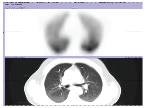
Figure 1: V/Q Scintigraphy in admission


Sehnaz Olgun Yildizeli1* Emel Eryuksel1 Fuat Dede2 Baran Balcan3 Berrin Ceyhan1
1Department of Pulmonology and Intensive Care, Medical School of Marmara University, Pendik Teaching Hospital, Turkey*Corresponding author: Şehnaz Olgun Yildizeli, Department of Pulmonology and Intensive Care, Medical School of Marmara University, Pendik Teaching Hospital, Pendik, Istanbul, Turkey, Tel: 902166452525; E-mail: drsehnazolgun@yahoo.com
Introduction: Percutaneous vertebroplasty is a vertebral augmentation procedure for treatment of painful vertebral fractures usually secondary to osteoporosis. Pulmonary embolism is a rare complication of this procedure and it usually remains undetected because of minor respiratory symptoms. In this report we report our experience with such a case.
Case report: A 48-year old male had spontaneous vertebral compression fractures secondary to osteoporosis. This was managed with percutaneous vertebroplasty was for each 11 vertebral body. On the first day after surgery, he described sudden onset of shortness of breath, chest pain and tachycardia.
Pulmonary embolism related to cement polymethyl methacrylate (PMMA) was diagnosed after diagnostic evaluation. The patient was treated with low molecular weight heparin (LMWH) and at the end of first week symptoms resolved. The procedure should be terminated if the patient became symptomatic or any leakage is detected.
Conclusion: Cement embolism is a complication of percutanous vertebroplasty procedures. Associated symptoms can occur during the procedure or postoperative period.
There is no specific standardized treatment protocol, but heparin therapy in emergency situation and warfarin in maintenance therapy are recommended. Anticoagulation may not be effective for resolution of the cement emboli in some cases, but may avoid additional thrombosis. Routine fluoroscopic imagining during and after procedure are suggested for early detection, using viscous cement material and performing venography may reduce the risk of embolism.
Vertebroplasty; Osteoporosis; Cement; Pulmonary embolism
Percutaneous vertebroplasty is a vertebral augmentation procedure for the treatment of painful vertebral fractures usually secondary to osteoporosis [1]. It is a relatively new procedure but becoming the standard care of pain control in these patients [2]. Polymethyl methacrylate (PMM) is used as cement in this procedure and main complications of the procedure are related with the leakage of cement. It can result in a wide range of complications as asymptomatic surrounding tissue damage, nerve root compression with persisting pain or pulmonary embolism [3-5]. Pulmonary embolism is a serious complication of these procedures.
A 48 years old man had a medical history of chronic obstructive pulmonary disease and diagnosis of relapsing uveitis but no proof of sarcoidosis or tuberculosis. He received pulse steroid and then oral steroid as maintenance for the treatment of uveitis. He had also smoking history for 30 years. He had spontaneous vertebral compression fractures secondary to osteoporosis. Percutaneous vertebroplasty was performed for each 11 vertebral body. First day after surgery patient presented with sudden onset shortness of breath, chest pain and tachycardia. Blood pressure was normal, heart rate was 116 per/min. lungs were clear to auscultation and percussion, cardiac and rest of the physical examination were normal. There were no specific changes in blood chemistry analyses and pH: 7.46, pCO2: 38 mmHg, pO2: 58 mmHg, HCO3: 28, SaO2: 91 in arterial blood gases analyses. A computed tomography CT angiogram of chest revealed a material denser from radiopaque, inside the hemiazygos vein of at right upper and lower lobes (Figures 1-3). Echocardiography and lower limb Doppler ultrasound were performed but there were no pathological findings. These results are consistent with cement PMMA pulmonary embolism and patient was anticoagulated with low molecular weight heparin LMWH and received oxygen therapy. One week later hypoxemia was resolved and patient discharged to home with LMWH treatment. Control CT angiogram revealed no change in PMMA embolism at the six month and ventilation perfusion scintigraphy (VQ) was performed. VQ scan also showed hypoperfused mismatch areas of right upper lobe anterior and left upper lobe apicoposterior but patient became asymptomatic after treatment (Figures 4 and 5).

Figure 1: V/Q Scintigraphy in admission
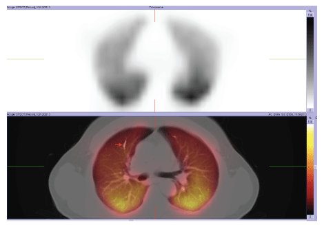
Figure 2: V/Q Scintigraphy in admission
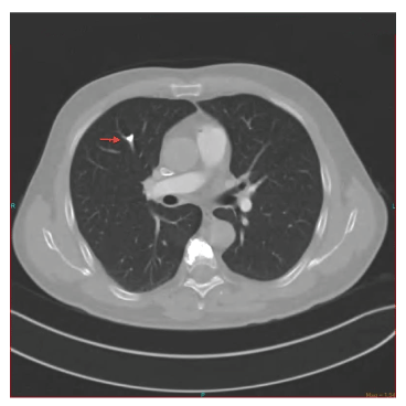
Figure 3: Thorax CT in admission
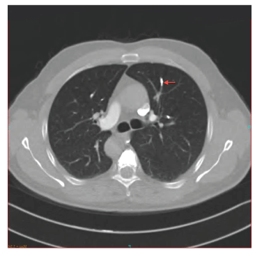
Figure 4: Thorax CT after LMWH Therapy
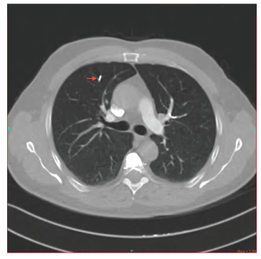
Figure 5: Thorax CT after LMWH Therapy
Percutaneous vertebroplasty is a minimally invasive fluoroscopic guided procedure usually preferred for the treatment of painful osteoporotic fractures and some vertebral invasive tumors [6]. PMMA is used as bone cement and injected to the vertebral bodies during the procedure.
Cement leakage to the surrounding tissue is the most common complication of the surgery [7] and reaches up to 90% [8]. Liquid consistency of PMM, injecting cement under higher pressure and treatment of malign tumors are related with the increased cement leakage complication rate [6]. In most of the cases leakage does not cause any problem and detected incidentally at X-Ray monitoring after surgery. Local PMMA leakage may cause nerve root compression, stenosis in the spinal canal, radicular pain and infection [9]. It also can be associated with some major systemic complications such as cerebral embolism, pulmonary embolism, renal artery embolism and acute respiratory distress syndrome [10].
Leakage of cement to the pulmonary vascular circulation from paravertebral veins is the main source of the pulmonary embolism. The incidence of cement embolism ranges to 3.5-23% and it tends to increase after vertebroplasty of osteoporotic fractures [11]. On the other hand there is a group of literature that reported no case of cement pulmonary embolism despite detected cement leakage into the venous system [12]. Clinically embolism can be symptomatic or asymptomatic. Symptoms are usually dyspnea, tachypnea, tachycardia, cyanosis, chest pain, hemoptysis but most of the patients can be totally asymptomatic or mildly symptoms can occur during the procedure or after a while from surgery. The incidence of patients presented with symptoms less than 1% in clinical practice [13]. Postoperative chest X-Ray usually detects pulmonary cement embolism as branching radio dense opacities, even though in clinically asymptomatic cases [9]. For further investigation CT angiography, ventilation perfusion scintigraphy echocardiography and lung diffusion capacity can be used to diagnose the embolism [5]. There is no standardized treatment of the cement embolism. Recommendations for the treatment are related to reduce the thrombus formation, pulmonary infarction and respiratory failure [9]. Localization, size of emboli also severity of the symptoms is effective on deciding the treatment choice [13,14]. Suggested treatment for symptomatic central orperipheral lesions is first intravenous heparin or LMWH after that warfarin or LMWH at least 3-6 months [13,14]. For severe symptomatic patients as in respiratory failure, embolectomy should be the first choice [14]. Anticoagulation may not reverse all cement emboli lesions but it is revealed that PMMA may lead to endothelial dysfunction and have prothrombotic effects that can result as additional thrombosis [15], so anticoagulation is essential to avoid from additional thrombosis or progressive occlusion of the pulmonary arteries. In elderly population continuous treatment after six month is not suitable because of the bleeding complications and side effects of the anticoagulation [6]. In the long term follow up patients may stay asymptomatic despite the consisting embolism.
In the literature there are several reports about cardiac perforation and tamponade secondary to cement embolism [16] and migratory intraatrial cement embolism [17] that all needed surgical procedure for treatment.
Review of the literature shows that this procedure should be performed by experienced surgeons under fluoroscopy. Viscous cement or toothpaste consistency reduces the leakage even though it is harder to work with viscous cement [18,19]. Also applying PMMA with a higher pressure especially at liquid consistency may also increase the leakage risk [6]. Patients should be monitored during the injection and procedure should be stopped immediately with detection of any leakage. Some authors advice routine fluoroscopic follow up for the first 24 hours after surgery [6]. Beyond these, prone positioning, performing venography before procedure, elevated intrathoracic pressure are suggested for the risk reduction [20,21].
Cement embolism is a complication of percutaneous vertebroplasty procedures. Symptoms can occur during the procedure or after postoperative period. Operation should be ended if patients become symptomatic or leakage detection during procedure. Routine fluoroscopic imagining during and after procedures are suggested for early detection using viscous cement material and performing venography may reduce the embolism risk.
Download Provisional PDF Here
Article Type: Case Report
Citation: Yildizeli SO, Eryuksel E, Dede F, Balcan B, Ceyhan B (2016) Non-Healing Radiologic Images of Cement Pulmonary Embolism after Percutaneous Vertebroplasty. J Clin Case Stu 1(5): doi http://dx.doi.org/10.16966/2471-4925.130
Copyright: © 2016 Yildizeli SO, et al. This is an open-access article distributed under the terms of the Creative Commons Attribution License, which permits unrestricted use, distribution, and reproduction in any medium, provided the original author and source are credited.
Publication history:
All Sci Forschen Journals are Open Access