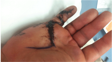
Figure 1: Post-operative appearance after removal of sutures at 2 weeks


Aparna Dasunmalee Ganhewa* Alex O’Beirne
Fremantle Hospital, Fremantle, Australia*Corresponding author: Aparna Dasunmalee Ganhewa, Fremantle Hospital, Fremantle, Australia, Tel: +61861617338; E-mail: dasun_ganhewa@hotmail.com
Here we present a case of a 26 year old man, who is a professional power lifter, engaging in regular weight lifting exceeding 300 Kg. During training a pop was felt in the right hand which resulted in an inability to flex at the distal interphalangeal joint of the right little finger. Although initially a Flex or Digitorum Profundus (FDP) avulsion type injury was suspected, intraoperatively a FDP Zone II tendon rupture was observed with a grossly abnormal tendon. Primary repair was undertaken. Post-operative compliance with hand therapy resulted in a normal functional outcome for the patient, with return to power lifting; now seven months post injury.
Closed Intratendinous ruptures of the flexor tendons of the hand secondary to trauma are rare. This is because of the strength of the tendon due to collagen cross linking. The usual points of detachment are at the musculotendinous junction, the muscle belly or at the point of tendon insertion with or without a bony avulsion fragment from the base of the distal phalanx.
Here we describe a case of spontaneous closed intratendinous Zone II, Flexor Digitorum Profundus (FDP) rupture in a professional power lifter. This case represents an interesting addition to the handful of cases currently reported with intratendinous Zone II FDP rupture to compare and contrast against.
This is an unusual case of a 26 year old man, right hand dominant and a professional power weight lifter. During his first repetition of dead lifts with 320 km weight, a “pop” was felt as he was holding the dead lift at the waist. Immediately the patient felt the right little finger unravels from gripping and there was loss of ability to flex the distal interphalangeal joint of his right little finger thereafter. Patient had pain and discomfort through the palm of the hand on flexion of his proximal interphalangeal joint (PIPJ). However flexion of PIPJ was intact.
Past medical history only included a raised body mass index (BMI) due to increased musculature, obstructive sleep apnoea requiring continuous positive airway pressure at night (CPAP) and fatty liver. At the time of injury the patient reported using a testosterone Enanthate for anabolic purposes. This was a twice weekly intramuscular injection of 500 mg of Testosterone Enanthate. At the time of injury a 10 week course had been completed. No past history of previous hand or finger fracture, no past history of rheumatoid arthritis or diabetes mellitus. Patient was a nonsmoker and had very minimal alcohol intake of approximately 2 standard drinks per year. No known drug allergies.
Our patient presented on the day of injury to the emergency department at Fiona Stanley Hospital in Western Australia. On examination, he had callused palms from weight training. The right hand and little finger were neuro-vascularly intact. No swelling or deformity noted. The volar aspect of the DIP was tender or palpation. Normal passive range of movement of both DIP and PIP joints. However there was no active flexion of DIP, and approximately 20 degrees flexion of PIP. Plain X-ray findings were unremarkable; particularly no avulsion fragment was seen from the base of the distal phalanx. The patient at this stage was diagnosed with a “Jersey finger” injury and discharged with an aluminum finger splint with a referral to the speciality hand surgery unit at Fremantle Hospital in Western Australia.
Our patient proceeded to theatre day 3 post injuries for exploration and repair of FDP under ultrasound guided regional forearm block and sedation. Pre-operative IV antibiotics were administered and a forearm tourniquet was used. The Little finger was opened with Brunner incision and extended to the palm. A zone II rupture of the FDP tendon was identified with the proximal end having retracted to the palm. A grossly abnormal and tendinopathic tendon was noted. This was colorfully described intraoperatively by the surgeon as a “tendon explosion” as the tendon fibres were torn and disrupted in a disorganized way. Unfortunately samples were not sent for histology at the time, or photos taken by medical imaging. The proximal and distal ends of the tendon were not trimmed to preserve length. The neurovascular bundles were visualized and protected. The tendon was repaired primarily with an interlocking whip stitch 2-0 Ti-Cron core suture and an interrupted 6-0 Prolene epitendinous suture. Intraoperatively there was good tendon glide and the finger was able to be fully extended and flexed. Skin closed with 5-0 Nylon and wounds dressed. A plaster of Paris dorsal blocking back slab was placed to prevent extension of the fingers (Figure 1).

Figure 1: Post-operative appearance after removal of sutures at 2 weeks
Post-operative management included placement into a dorsal blocking thermoplastic splint and very gentle range of movement was commenced as per protocol day three post-op. The Fremantle hospital hand unit adopts a protocol similar to the Belfast flexor tendon protocol. Hand therapy included warming with wax, active and passive range of movement, tendon gliding exercises, place and holds and scar massage. Beige putty was used at week 5 to provide small amount of resistance while forming a fist and hook with the hand. The initial five weeks of hand therapy showed little gain in active flexion. At week five, patient had 0-4 degrees of active flexion and an ultrasound was performed at this stage to ensure that the repair was intact and the tendon was in continuity, which it was. With extreme compliance and diligence with the hand therapy regime, active flexion gradually returned over the next three to four weeks. Week 6 active flexion at DIP 0-8 degrees, week seven: 0-24 degrees, week 8: 0-36 degrees, week nine: 0-52 degrees (Table 1).
| Week post operatively | Active Range of Movement of right little finger DIPJ (extension-flexion) in degrees |
| Week 5 | 0-4 |
| Week 6 | 0-8 |
| Week 7 | 0-24 |
| Week 8 | 0-36 |
| Week 9 | 0-52 |
Table 1: Active Range of Movement of right little finger DIPJ (extensionflexion) in degrees
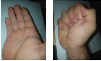
At the time of discharge at week 12, there were no flexion deformities involving either PIP or DIP, active range of movement was 0-96 at MCPJ, 0-76 at PIPJ and 0-56 degrees at DIPJ. However, currently 11 months post operatively a 20 degree flexion contracture has developed involving the PIPJ. Patient has since been re-referred to continue with hand therapy (Figure 2).

Figure 2: Range of movement 7 Months Post Injury
Intratendinous tendon rupture involving the flexor tendons of the hand are rare, furthermore Zone II ruptures are rarer still. The literature review done by Bios et al. [1] found, out of the total number of known cases in the literature at the time in 2006, seven cases involved a Zone II. This review found the most common zone of injury for rupture is zone III (80%) and the most common site of rupture in zone III is at the origin of the lumbricals [2,3,1]. The next most common site of rupture is Zone II (14%), followed by Zone IV (6%).
In the literature, reported cases of Zone II rupture include the one as described by Naohito et al. [4], of a man falling down stairs resulting in rupture of FDS and FDP. Other cases include one case in the series presented by Naam et al. [2], and 2 cases in the series presented by Imbriglia et al. [5]. The case by Prosser et al. [6] is one of FDP rupture in proximal zone II at the level of the A1 pulley. An acute tenosynovitis was seen in this case. In the case presented here the tendon was discovered to be grossly tendinopathic and it would have been useful to have a histological correlation to these intraoperative findings. No samples were however sent for histology.
It is interesting to compare and contrast the FDP avulsion (jersey finger injuries) with FDP intratendinous rupture. For example the most commonly affected finger in avulsions is the ring finger, however with rupture it is the little finger. A reason for the vulnerability of the little finger FDP may be due the importance of the little finger for grip strength, the load through little finger FDP is greater than other fingers. Furthermore it is reported that 34% of general population have an absence of FDS to the little finger [1].
Similar to the FDP avulsion injuries occurring at the FDP insertion point, ruptures are most commonly caused by forced extension against a maximally flexed finger or flexion against resistance [2,7]. It is acceptable knowledge that tendons do not generally rupture unless there is an underlying pathological process. McMaster et al. [8] showed experimentally that the tendon needs to lose >50% of its tensile strength before rupture occurred. It is suggested that intratendinous rupture occurs nearly always in the context of a pathologic tendon. However the exact pathogenesis of spontaneous tendon rupture in the hand remains unclear. Certainly in the case described here rupture occurred with load and flexion against resistance and intraoperatively the tendon was grossly abnormal.
McLain et al. [9] suggested an intratendinous rupture occurs due to atypical loading or strain in the context of chronic vascular compromise or repeated micro trauma. A relative region of avascularity is suggested to exist in 2 areas of the tendon: a watershed area in zone 2, and also at the insertion of the lumbricals. It is entirely plausible that the cause of rupture in our case described is due to chronic vascular comprise as well as repeated micro trauma caused by repeated loading with regular weight lifting.
In conclusion we are pleased to present a further case to the literature of a Zone II FDP tendon rupture. Furthermore it is an interesting case of a young man who is a professional power lifter. This case had a very favourable outcome with the patient proceeding to continue with power lifting up to 360 kg 7 months post repair.
Download Provisional PDF Here
Article Type: Case Report
Citation: Ganhewa AD, O’Beirne A (2016) An Unusual Case of Zone 2 FDP Tendon Rupture in a Professional Power Lifter. J Clin Case Stu 1(4): doi http://dx.doi.org/10.16966/2471-4925.127
Copyright: © 2016 Ganhewa AD, et al. This is an open-access article distributed under the terms of the Creative Commons Attribution License, which permits unrestricted use, distribution, and reproduction in any medium, provided the original author and source are credited.
Publication history:
All Sci Forschen Journals are Open Access