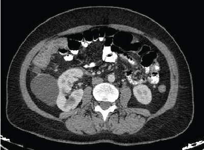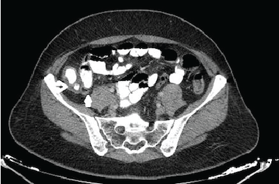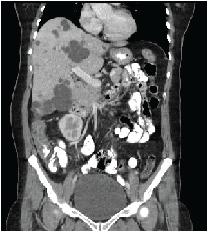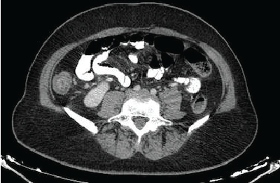
Figure 1: Axial abdominal CT with IV and oral contrast-Open arrow: Transmural concentric wall thickening of caecum/Thick arrow: Normal appendix vermiformis


Ceren Yalnız1 Kaan Meriç1* Hasan Gündoğdu1 Mehmet Fatih Aydoğan2 Mehmet Kamil Yıldız3 Hacı Hasan Abuoğlu3 Mehmet Masum Şimşek1
1Haydarpaşa Numune Eğitim ve Araştırma Hastanesi, Radyoloji Kliniği, İstanbul, Turkey*Corresponding author: Kaan Meriç, Haydarpaşa Numune Eğitim ve Araştırma Hastanesi, Radyoloji Kliniği, İstanbul, Turkey, Tel: 00902163360565; E-mail: drkaanmeric@hotmail.com
Neutropenic enterocolitis is a serious complication of neutropenia following the administration of antineoplastic and immunosuppressive drugs. It was mostly observed in patients with hematologic malignancies in the past. However, after the increased use of chemotheraphy agents and immunosuppressive therapy, it has been reported more frequently. We present a patient with rheumatoid arthritis (RA) presented with neutropenic enterocolitis induced by methotrexate therapy.
Computed tomography; Methotrexate; Neutropenic enterocolitis; Rheumatoid arthritis
Neutropenic enterocolitis is defined as transmural bowel wall inflammation,which may cause perforation and sepsis in patients with severe neutropenia. It is most frequently seen in neutropenic oncologic patients treated with cytotoxic chemotherapy agents. Aplastic anemia, myelodisplastic syndrome and acquired immune deficiency syndrome are notable non-oncological conditions associated with neutropenic enterocolitis.
In this case report, we present a RA patient who developed neutropenic enterocolitis associated with methotrexate treatment.
A 47-year-old woman presented to our emergency room with fever, right lower abdominal pain, nausea and vomiting. During ultrasound imaging performed to rule out acute appendicitis, diffuse caecum wall thickening was noted but the appendix vermiformis could not be visualised. Intravenous and oral contrast-enhanced computed tomography of the abdomen demonstrated circumferential wall thickening of caecum (Figures 1 and 2), ascending (Figures 2 and 3) and transverse colon (Figure 4), measured 11 mm at most. A few paracolic lymph nodes were also noted, the largest with a short axis-diameter of 5 cm. The localisation, size and shape of appendix vermiformis were all normal (Figure 1).

Figure 1: Axial abdominal CT with IV and oral contrast-Open arrow: Transmural concentric wall thickening of caecum/Thick arrow: Normal appendix vermiformis

Figure 2: Coronal abdominal CT with IV and oral contrast-Transmural concentric wall thickening of caecum and ascending colon

Figure 3: Axial abdominal CT with IV and oral contrast-Transmural concentric wall thickening of ascending colon

Figure 4: Axial abdominal CT with IV and oral contrast-Transmural concentric wall thickening of transverse colon
The laboratory findings revealed that the patient had pancytopenia.The patient had RA and was receiving methotrexate therapy for more than ten years.Our final diagnosis was neutropenic enterocolitis associated with methotrexate induced pancytopenia.
Neutropenic enterocolitis, also known as typhlitis, is an important complication of neutropenia associated with malignancy and chronic inflammatory diseases. Although the pathogenesis of neutropenic enterocolitis is not exactly understood, mucosal damage caused by cytotoxic drugs or neutropenia might be the predisposing factor. It is thought to result from the microbial invasion of damaged intestinal mucosa,leading to bowel wall edema and inflammation [1]. It can rapidly progress to intestinal perforation, multisystem organ failure, and sepsis. Presenting signs and symptoms are fever, abdominal pain, nausea, vomiting, and diarrhea. Rapid diagnosis and aggressive medical and/or surgical intervention are extremely important for the mortality of the patient [2-3].
Neutropenic enterocolitis is a rare complication of medical therapy for RA, and only two cases have been reported in literature. In 1992 Chakravarty et al. [4] described the first case of neutropenic enterocolitis in a 69-year-old RA patient following sulphasalazine therapy. The second case was reported by Lin et al. in 2014. [5]. They reported a patient with RA presented with neutropenic enterocolitis as a complication of methotrexate therapy
Since neutropenic enterocolitis is a rare but life threatening complication of methotrexate therapy, it should always be on the differential diagnosis list of acute appendicitis, diverticulitis, abcess and inflammatory bowel diseases. When computed tomography of the abdomen demonstrates circumferential bowel wall thickening, intramural oedema or haemorrhage and pericolic inflammation in a patient with neutropenia, the most likely diagnosis is neutropenic enterocolitis, which does not warrant further work up or surgery [6].
Neutropenic enterocolitis is a rare but highly dangerous complication of neutropenia from any causes.It occurs mostly in oncology patients and patients with chronic inflammatory diseases,who receive high doses of cytotoxic chemotherapy drugs or immunosuppressive drugs. A neutropenic patient presenting with symptoms such as fever,abdominal pain,vomiting and diarrhea should be evaluated for neutropenic enterocolitis for early diagnosis and appropriate therapy.
Download Provisional PDF Here
Article Type: Case Report
Citation: Yalnız C, Meriç K, Gündoğdu H, Aydoğan MF, Yıldız M, et al. (2016) Neutropenic Enterocolitis Associated with Methotrexate Toxicity. J Clin Case Stu 1(5): doi http://dx.doi.org/10.16966/2471-4925.126
Copyright: © 2016 Yalnız C, et al. This is an open-access article distributed under the terms of the Creative Commons Attribution License, which permits unrestricted use, distribution, and reproduction in any medium, provided the original author and source are credited.
Publication history:
All Sci Forschen Journals are Open Access