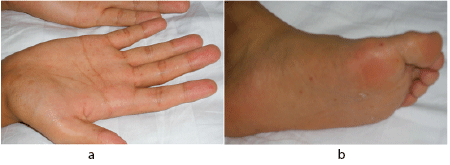
Figure 1: a) HFMD: vesicular rash on palms. B) HFMD: vesicular rash on feet

Rui Song1* Wenhao Hua1,2 Zhihai Chen1 Xingwang Li11
1The National Clinical Key Department of Infectious Disease, Beijing Ditan Hospital, Capital Medical University, Beijing, China*Corresponding author: Rui Song, The National Clinical Key Department of Infectious Disease, Beijing Ditan Hospital, Capital Medical University, Beijing, China, Tel: +0086 13126595640; E-mail: songruii@hotmail.com
Hand, foot, and mouth disease (HFMD) occurs frequently in children. Generally, most of HFMD cases are mild, while severe neurological and systemic syndromes occurred in some patients as well. This 20-year-old man from Beijing infected with HFMD and also showed the severe neuro-symptom is rarely seen before. He presented with an acute, self-limiting illness characterized by high temperature and vesical rashes eruptions on his hands, feet, and mouth. Serologic and RT-PCR tests indicated that his illness was associated with Coxsackie virus A16 (CVA16). In addition to typical clinical signs of HFMD, the patient presented with visual field defects. Notably, magnetic resonance imaging (MRI) examination indicated the lesions in his brain. In addition to the typical clinical signs, visual field defects and lesions in brain occurred in the adult with CV-A16 infection.
Hand, Foot and Mouth disease; Coxsackie virus A16; Adult; Visual field defects; Brain lesions
Hand, foot, and mouth disease (HFMD) is caused by a group of human enteroviruses (HEV). It’s typical clinical manifestations include fever, rashes and vesicles on the hands, feet, and mouth. HFMD occurs mainly in children, particularly in those less than 5 years of age [1]. Most of HFMD cases were mild and limited to fever and vesicular rash on palms, soles, and mouth, while severe neurological and systemic syndromes that can be fatal occur in some patients, particularly in those caused by enterovirus 71 (EV71) [2]. HFMD occurs rarely in adults, but a few of cases have been reported to exhibit myocardial damage [3], lung infection [4], respiratory failure [5] and neurological symptoms such as nausea, vomiting, and decreased muscle strength [6].
EV71 and Coxsackie virus A16 (CV-A16) are the most common agents of HFMD in China [7]. Although adult HFMD cases caused by EV71 and CV-A16 were reported [8], no cases have been associated with severe neurological and systemic syndromes. Even in children cases, they seldom show the symptom of visual field defects and image lesions. Here we report a severe adult HFMD case caused by CVA16, with acute visual field defects, headaches, and other neurological symptoms. Also, an MRI scan was taken that indicated some lesions on the brain. The image of MRI also showed the lesion in his brain.
In August 2013, a 20-year-old male resident of Haidian District, Beijing, was hospitalized in Beijing Ditan Hospital with a 6-day history of fever and rash on his hands, feet, and oral ulcers. Remarkably, his visual acuity decreased from the third day of onset. The blurred vision became worse, almost with visual field loss on the fourth day of onset. Additionally, he felt severe headache without vomiting. The patient had been healthy previously, and there was no history of exposure to patients with similar syndromes.
Blood cultures for anaerobes and aerobes were all negative on 3 different occasions. Throat swabs and fecal samples were collected for pathogen test. The broad range RT-PCR screen for enteroviruses including EV71, CVA16, and Echo virus was performed. As a result, only RT-PCR targeting to CVA16 showed positive. Serological test was strongly positive for immunologlobulin (M) to CVA16 antigens. Additionally, serological assays and PCR were performed to detect other pathogens including herpes virus. All these tests were negative. These data indicated that the pathogen was CV-A16.
Upon admission, general physical examination for the patient showed: high temperature (38.6°C), BP of 104/66 mm/Hg, and pulse rate of 110 beats/min. The patient presented with scattered erythematous macules and grayish oval vesicles, with 2 to 4 mm in size, on the hands and feet (Figure 1). There were small vesicles on the upper palate. He had mild neck stiffness. But his muscle strength, muscle tone, and tendon reflexes of the limbs were normal.
Figure 1: a) HFMD: vesicular rash on palms. B) HFMD: vesicular rash on feet
His visual fields had defects. Ophthalmological test showed as follows: his eyesight were : right eye 0.25, left 0.6; clear cornea, deep anterior chamber, round pupil, reactive to light, clear lens, no fundus abnormality after mydriasis for both eyes. The erimetry test revealed: homonymous hemianopsia (upper left) for both eyes, hence he was diagnosed as hemianopsia (homonymous hemianopsia of both eyes), accompanied by bilateral ophthalmalgia.
Laboratory tests were the following: white blood cell (WBC) 14.44 × 10⁹/L (4-10 × 109/L), neutrophil 85.54% (50%-75%), c-reactive protein (CRP) 88.89 mg/L (0-10 mg/L), procalcitonin (PCT) <0.05 ng/ml (0-0.05 ng/ml), CD4+ T cells 763 cell/µl (706-1125 cell/µl). Auto antibodies were all negative. Cerebrospinal fluid (CSF) examination revealed: pressure 210 mmH2O (80-180 mmH2O), total cell counts 70/µl, leukocyte counts 40/µL (0-5/µL), protein 44.60 mg/dL (8-43 mg/dL), Glucose 4 mmol/L (2.5-4.5 mmol/L), Cl 118.80 mmol/L (120-132 mmol/L). The CSF showed the virus infectious meningitis. In addition, cerebral magnetic resonance imaging (MRI) demonstrated the cortical and subcortical white matters lesions on the right occipital lobe. Lesions showed slight density enhancement. This image suggested the occurrence of new infarct (Figure 2).
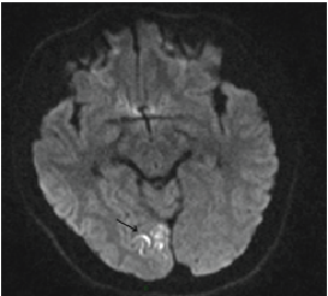
Figure 2: Head MRI of the case
The arrow shows the local lesion: signals in the cortex and sub cortical white matter of the right occipital lobe, possibly new infarct
During hospitalization, mannitol was used for dehydration and relief treatment for other symptoms. No steroids or antivirus drugs were used. Two days later after admission, the blurred vision went into remission spontaneously and the visual field recovered as well. The patient was discharged one week later and with no other complications.
Although HEV infection in children presents the obvious clinical syndrome (HFMD), the infection in adults is often latent or only presents mild symptoms [9]. The typical clinical features including skin lesions and pustular rash are only observed in a few of adult infections [10]. Sporadic cases presented with headache, nausea, pulmonary symptoms, arthralgia, and arthritis [5]. To date, only one CV-A16 infection with fatal pneumonitis in a 76-year-old man has been reported [11]. Here, to our knowledge, it is the first case of CV-A16 infection in adults exhibited HFMD symptom with headache, nausea, and admitted to hospital for the visual field defects.
The case’s CSF examination revealed that elevated cell counts and mildly increased protein level, consistent with viral encephalitis. His visual symptoms were cooperating with the lesion in the occipital lobe as showed in MRI. Bartels [12] reported, infarction in the occipital lobe accounts for 3–6.4% of all cerebral infarctions. The occipital lobe is the visual center in the cortex, and each field of the retina projects to a certain position in the striate cortex at the occipital lobe. For this reason, when occipital lobe infarction occurs, there are changes in the visual field [13]. There are many causes of occipital infarction, but occipital infarction in association with CVA16 infection has not been previously reported. Whether the neurological symptoms and visual field defect of the patient are a result of impaired central nervous system due to immune responses caused by acute viral infection is still not clear, and there has been no relevant literature.
The patient recovered and didn’t receive any special medicine, which showed it maybe a self-limited course. Additional, similar cases must be evaluated and analyzed to determine why such a unique clinical manifestation occurred in this patient, whether it was related to the virus or due to individual variation. We should do more surveillance on the adult HFMD.
This study was supported by “the capital health research and development of special (2014-4-2175)”.
Download Provisional PDF Here
Article Type: Case Report
Citation: Song R, Hua W, Chen Z, Li X (2016) A Case of Severe Hand, Foot and Mouth Disease Accompanied by Visual Field Defects in Adult Patient. J Clin Case Stu 1(6): doi http://dx.doi. org/10.16966/2471-4925.106
Copyright: © 2016 Song R, et al. This is an open-access article distributed under the terms of the Creative Commons Attribution License, which permits unrestricted use, distribution, and reproduction in any medium, provided the original author and source are credited.
Publication history:
All Sci Forschen Journals are Open Access