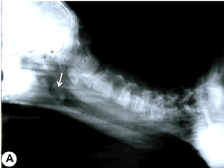
Figure 1: Plain x-ray film in a latero-lateral view of the cervical region shows the pharyngeal part of the esophagus filled with air (white arrow)

Magda M Ali Ahmed Ibrahim*
Department of Surgery, Anesthesiology and Radiology, Faculty of Veterinary Medicine, Assiut University, Assiut; Egypt*Corresponding author: Ahmed Ibrahim, Faculty of Veterinary Medicine, Assiut University, Assiut, Postal Code: 70155, Egypt, Tel: +201062204009; Fax: 088-208050; E-mail: elgrah38@gmail.com
A case of two months old cattle calf was admitted to the veterinary teaching hospital, Assiut University, Assiut, Egypt. The animal was suffering from difficulty in swallowing with regurgitation of milk after suckling since one and half month. In addition gaseous distension of the abdomen and dehydrated skin. Clinical examination revealed the presence of a 10 cm long soft, cervical swelling on the left side of the neck, with tympany. Passage of the stomach tube was not possible at the level of the mid way of the esophagus. Plain and contrast radiography of the esophagus showed an esophageal dilatation followed by a stricture at the level of the midway of the esophagus. Surgical exploration of the esophagus, showed a dilated part from the esophagus followed by a stricture with a cross sectional membranous obstruction. The animal was treated surgically and the membrane was removed; thereafter, the passage of the stomach tube was possible.
Esophagus; Obstruction; Cattle calf
Esophageal stricture is a pathologic narrowing of the lumen that may becomes the main cause of swallowing difficulties and usually develops after trauma (eg, foreign body), ingestion of caustic substances, exposure to certain drugs, gastro esophageal reflux, or tumor invasion [1,2]. It is also linked with major diseases of gastrointestinal tract such as dysfunctional lower esophageal sphincter, hiatal hernia, disordered motility and esophagitis. Most strictures develop in the thoracic portion of the esophagus. Idiopathic esophageal strictures are recorded in foals. Initial diagnosis based on clinical signs may be delayed because of other more frequent causes of dysphagia, including idiopathic dorsal displacement of the soft palate or nasal reflux of milk, cleft palate, or pharyngeal cysts [3]. The primary indication for esophageal surgery in large animals is to relieve esophageal obstructions, which have not responded to conservative treatment. Surgical exploration of the esophagus in the bovine is not commonly indicated and frequently cannot be justified on an economic basis [4].
Two months old cattle calf was presented to the veterinary teaching hospital of faculty of veterinary; Assiut University; Assiut; Egypt; with the case history of difficulty in swallowing with regurgitation of milk after suckling since one and half month, the owner reported that the animal was normal in suckling after birth and the symptoms began after giving the animal horse bean. In clinical examination, the calf was relatively in a fair health condition, but the skin appeared dehydrated. The neck was stretched, rectal temperature, respiratory rate and pulse rate were normal; a clear cervical swelling of about 10 cm length on the left side over the esophagus. The swelling was soft and non painful. In addition, tympany was observed on the abdomen. Attempts to test the passage of water through a stomach tube failed and the water was regurgitated mixed with milk and saliva.
Oral introduction of stomach tube failed and the passage of the stomach tube was stopped at the lower level of the swelling. During removal of the stomach tube, its distal end was filled with milk and saliva indicating that the swelling is filled.
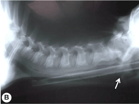
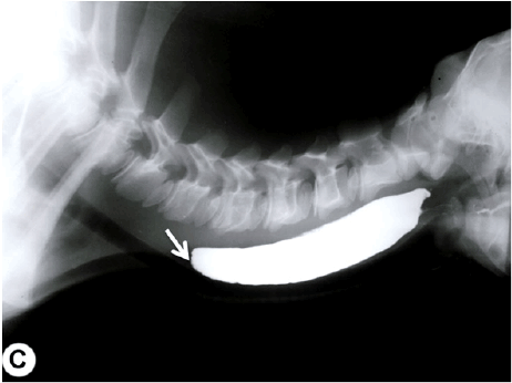
Plain x ray was taken for the cervical region in a latero‐lateral view with and without the presence of the stomach tube (Figures 1 and 2). Contrast radiographic examination was performed using a suspension of barium sulfate (60% w/vol.) as a contrast agent that was given through the stomach tube also in a latero‐lateral view.
The majority of the contrast medium was returned back from the mouth cavity. The contrast x ‐ray film showed the retention of the barium inside the swelling without any escape of the barium down word the lower level of that swelling (Figure 3). According to the previous findings, a decision was made to perform an exploratory esophagotomy.

Figure 1: Plain x-ray film in a latero-lateral view of the cervical region shows the pharyngeal part of the esophagus filled with air (white arrow)

Figure 2: Plain x-ray film in a latero-lateral view of the cervical region shows stomach tube inside the esophagus, the end of the stomach tube is the area of obstruction, notice the dilated part of the esophagus filled with air (white arrow)

Figure 3: Contrast x‐ray film in a latero-lateral view of the cervical region shows the contrast medium inside the esophagus, the radio paque area is the dilated part from the esophagus. The white arrow indicates the seat of the membranous obstruction
The calf was restrained on the right side and the surgical site over the dilated swelling at the level of the med way of the neck was routinely prepared for surgery.
Under the effect of local anesthesia using 1% Lidocaine Hcl; a 10 cm long incision was made passing through the skin and subcutaneous tissues, sharp and blunt dissection between the Sterno‐cephalic muscle and the trachea allowed approach of the esophagus. About 10 cm long dilatation in the esophagus was clear at the med way of the esophageal length followed by normal size of the esophageal lumen. Insertion of the stomach tube was repeated with massage on the outer surface of the esophagus without success. The operation site was packed with sterile towels to avoid any contamination by fluid or ingesta from the esophagus.
A longitudinal incision was made over the dilated part as well as the strictured point. The dilated part was filled with remnants from the contrast medium. In exploration of the esophagus, a thin transverse membrane was seen closing the end of the dilated part with a very narrow opening at the middle of the membrane allowing passage of scanty amount of fluids through it, the remaining lumen behind the membrane was slightly stenosed. The membrane was incised and removes carefully without injuring the esophageal mucosa and the stomach tube was reintroduced to allow rupture of the remaining parts from the membrane and to examine the free passage of it through the whole esophageal length. The dilated part from the esophagus was not removed and the esophageal mucosa was sutured using simple continuous suture pattern with chromic cat gut no. 0, the submucosa and muscular layer were closed as a one layer in the same manner. The subcutaneous tissue was closed with simple continuous pattern with chromic cat gut no. 1 and the skin was closed with horizontal matters suture pattern using Silk no. 2.
Postoperative care included intramuscular injection of antibiotic and fluid therapy for three days. The calf was first allowed to fed normal four days post operatively. The stitches were removed 10 days after operation. The animal was able to drink after surgery but some of the milk was also.
Initial diagnosis of esophageal stricture based on clinical signs may be delayed because of other more frequent causes of dysphagia, including idiopathic dorsal displacement of the soft palate or nasal reflux of milk, cleft palate, or pharyngeal cysts [5]. The clinical signs are similar to those associated with foreign bodies and include regurgitation, ptyalism, dysphagia, and pain. In our examined case, the clinical signs were similar to those reported before and were not specific for stricture. The use of radiography gives the best result especially with the use contrast material as Barium sulfate. Survey and contrast radiographs are recommended to characterize lesions in the bovine esophagus. The use of contrast radiography allow exact determination of the seat of stricture and the degree of dilatation of the esophagus [6,7]. The technique is inexpensive, do not need special precautions and could be easily applied. Other authors reported that an esophagram under fluoroscopy is the preferred tool for diagnosis, because it allows visualization of the number, length, location, and severity of strictures [8,9]. This technique was not used in our study to avoid the high radiation hazards accompanying Floroscopy. Esophagoscopy can also be diagnostic but does not allow visualization beyond the stricture unless esophageal balloon dilation is also performed [10]. In addition, the use of endoscopy is expensive and need good experience.
Most strictures were reported to develop in the thoracic portion of the esophagus [5]. This was not the seat for stricture in our study as the area for stricture was at the med way of the cervical part of the esophagus. Esophageal stricture in horses or ruminants typically results from mucosal ulceration secondary to esophageal obstruction [11]. The cause for stricture in our case is not clear but congenital cause could not be accepted as according to the case history, the animal was suckling normally since birth and till the end of the first month of age. As the results began to occur after giving the animal horse bean in this young age; therefore the condition could be secondary to esophagitis or mucosal ulceration. Appropriate treatment depends on whether the stricture is mucosal or mural (involving the muscular wall). Mucosal strictures can be treated conservatively with dietary management, bougienage with a cuffed endotracheal tube, or surgery [5]. In our studied case, esophagotomy was indicated to determine the main cause for stricture and to resect the membranous obstruction which prevents passage of milk and food into the esophagus. Although the primary indication for esophageal surgery in large animals is to relieve esophageal obstructions, which have not responded to conservative treatment, surgical exploration of the esophagus in the bovine is not commonly indicated and frequently cannot be justified on an economic basis [5]. In our study, no conservative treatment was helpful and resection of the membranous obstruction to reinforce the normal movement in the esophagus could only be performed via esophagotomy. Restriction of oral feed and water postoperatively is important to help prevent breakdown of the suture line in the esophageal wall and subsequent fistula formation [11]. This was also performed in our study and the animal was supported with fluid therapy to prevent dehydration and the animal was allowed to drink milk and water from a bottle.
The presence of membranous obstruction in the esophagus could be secondary to esophagitis after false feeding protocols. Esophageal dilatation in front of the membrane due to accumulation of milk and food particles leads to weakness in the esophageal movement and regrution of food in addition to formation of an esophageal stricture behind the obstructed area. Surgical treatment is the only way to treat such cases.
Download Provisional PDF Here
Article Type: Case Report
Citation: Ali MM, Ibrahim A (2016) Successful Surgical Management of Cervical Esophageal Membranous Obstruction and Stricture in a Cattle Calf. J Clin Case Stu 1(1): doi http://dx.doi. org/10.16966/2471-4925.105
Copyright: © 2016 Ali MM, et al. This is an open-access article distributed under the terms of the Creative Commons Attribution License, which permits unrestricted use, distribution, and reproduction in any medium, provided the original author and source are credited.
Publication history:
All Sci Forschen Journals are Open Access