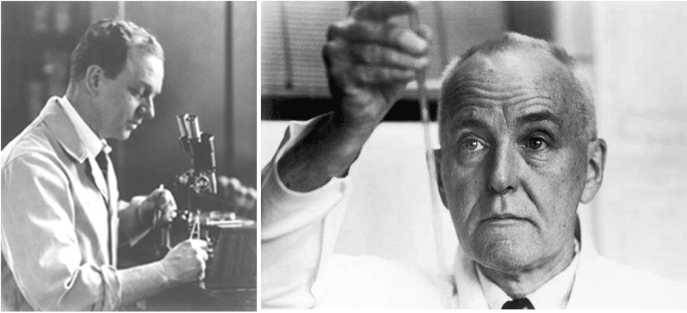
P. Rous a few years after the discovery of RSV. C. Huggins working in the laboratory

Ugo Rovigatti*
Department of Medicine, University of Florence, Italy
*Corresponding author: Ugo Rovigatti, Department of Medicine, University of Florence, Viale Pieraccini 6, 55139, Florence, Italy, Tel: +39-3895608777; E-mail: profrovigatti@gmail.com
Aritcle Type: Editorial
Citation: Rovigatti U (2016) The Lesson of Molecular Oncology and Prostate Cancer: Celebrating 50 years of the Nobel Award to P. Rous and C. Huggins. Int J Cancer Res Mol Mech 3(1): doi http://dx.doi. org/10.16966/2381-3318.e102
Copyright: © 2016 Rovigatti U. This is an openaccess article distributed under the terms of the Creative Commons Attribution License, which permits unrestricted use, distribution, and reproduction in any medium, provided the original author and source are credited.
Publication history:
In the fast-pace of Cancer Research (CR) activities, high throughput technologies and accumulation of data by NGS and –OMICS in general, some milestones in our understanding of cancer may easily skip our attention. This year marks 50 years from the assignment of two Nobel Prizes for CR in 1966: to Peyton Rous and Charles Huggins [1-4]. Some reflections may be worthier, since they also impinge on today’s state of affair in CR. First of all, the two certainly appeared as an “odd couple” in 1966 [5]. A celebrated virologist and immunologist from the Rockefeller University (Institute at that time), the work of Rous -87 at that timeseemed to have little to share with the clinical breakthroughs just realized by the Canadian endocrinologist Huggins at the University of Chicago. Although coming from widely different areas of medicine (microbiology and surgery) they both knew and highly respected each other [2]. Furthermore, these two CR giants certainly overlapped in their belief that research intuition and scientific ingenuity and discovery would be at the basis of any further development in science.
Rous discovery was made the same year he entered Rockefeller (1909), when a hen of a particular breed of Plymouth was brought to his attention from a farmer in view of a prominent wing sarcoma. Not only could he transfer the tumour with isolated cells, but also with a cell-free extract and after ultra-filtration, thus excluding transfer of entire cells (or even bacteria) and strongly implicating presence of an infecting virus. The initial paper was published in 1911 by the Journal of Experimental Medicine and followed by several additional reports characterizing the system [6-7]. However, Rous group could never isolate the responsible virus, now called RSV, and finally shifted in 1915 to other relevant biomedical subjects of research. Still, he returned to viral oncology in the 1930’s by studying a different virus: the Papilloma Virus in rabbits, that Shope had isolated a few years earlier [8]. Only several years later, starting in the mid 50s, a systematic study of RSV tumorigenesis in vivo and transformation of chicken cells in vitro was initiated in the laboratory of R. Dulbecco in California and with great pioneers such H. Rubin, H. Temin, H. Hanafusa, P. Vogt, P. Duesberg, M. Bishop, H. Varmus, R. Weiss, J. Svoboda etc. (for excellent reviews of this field, see [9,10]). This initially led to the discovery of reverse transcriptase by Temin’s and Baltimore’s laboratories and finally to the isolation of the first oncogene, baptised SRC, from RSV viral genome in the laboratory of Bishop-Varmus: also these discoveries were awarded by the Nobel Prize (in 1975 and 1989 [11,12] ) and most of all the birth of a new era called molecular oncology.

P. Rous a few years after the discovery of RSV. C. Huggins working in the laboratory
The seminal discovery of C. Huggins was published 30 years later in 1941, when he reported with colleague Clarence Hodges that androgen ablation -either by surgical castration or by giving estrogens (stilbestrol) as androgen antagonists- causes prostate cancer (PCa) atrophy and regression. Starting from the first attempt, this approach has been very successful, since it allowed patients with metastatic PCa to immediately experience great pain relief and amelioration due to tumour shrinkage and final atrophy [3-4]. Today, we know that PCa can be a rather indolent disease, therefore controllable by Androgen Depletion Therapy (ADT). Unfortunately, in many cases ADT or surgical castration is followed by metastatic castration resistant prostate cancer (mCRPC), a condition for which we don’t have today curative solutions [13]. To conclude the parable about the odd couple in 1966 Nobel sharing, the following half Century has shown how the two discoveries have much assonance, and yet they present today common difficulties or open questions [5,14,15].
What we call Molecular Oncology was created from the seminal discovery of P. Rous in 1911 and the patient ingenuity of H. Varmus and M. Bishop several years later (among many others) who finally provided a rational interpretation of the molecular intricacies of cancer cells. The most acclaimed of such new molecular interpretations is the one proposed as “Hallmarks of Cancer” (HoC) by D. Hanahan and R. Weinberg in 2000 and 2011 and as an even more detailed “mosaic” of every aspect of molecular-cancer in the 2014 book “The Biology of Cancer” [16-18]. Unquestionably, such molecular reform started from Rous seminal intuition, had the great merit–both historical and epistemological- to put under the same umbrella the many disparate pathologies (at least 50 of them), that we call cancer. Yet and as later discussed, it is clear today that also this approach or model should be revised [14,15].
As previously discussed, today we can progress at much faster pace in view of accelerated technological advances in the so called NGS Era [15], although a detailed description of cancer in general- and PCa specificalterations had been initiated also in the pre-NGS era [19]. Since essentially every human cancer has been affected by this kind of revolution, what are today’s implications for Huggins discoveries and PCa in particular? 1. PCa sets itself apart from other neoplasms since it shows the lowest number of point-mutations or SNVs (single nucleotide variants) among human cancers (in the order of .33/1.4 per MegaBase) [20,21]. This already contradicts a long-accepted tenet of molecular oncology: that cancer may be just caused by accrual/accumulation of point mutations in essential regulators of cell-cycle/cell-control (so called cancer-drivers) [14,22,23]. Interestingly, it also agrees with the consistently and almost stubbornly defended opinion of Rous that point-mutations have little relevance in cancer onset and progression [2,7]. 2. Lack of point mutations is amply compensated in PCa by extensive presence of variations in copy numbers (CNAs): among amplifications, MYC and AR genes are the most frequent, while several regions believed to contain TSGs (tumor suppressor genes) are also often deleted (NKX3, PTEN, CDKN1B, RB1, TP53) [24]. 3. Major alterations of PCa genomes appear to be dichotomized, as approximately 50% contain rearrangements between the TMPRSS2 gene (androgen responsive) and transcription factors of the ETS family (E26 Transformation Specific), while the other 50% typically contain deletions of CHD1 or mutations in SPOP1 [20,24-26]. 4. One of the major problems of PCa results, is the very strong tumour heterogeneity they have demonstrated. For example, only two overlapping genes were mutated in >15% of cases in two extensive cohorts of patients studied by exome sequencing (inter-tumor heterogeneity) [20]. Two studies have also demonstrated very extensive intra-tumor heterogeneity: in ¾ of patients, Lindeberg et al. [27] found no common somatic mutations in different nodules of the same PCa patient and Boutros et al. [28] demonstrated also no common CNA in different foci from the same patients.
The very rapid evolution of the past 50 years has essentially vindicated the close relatedness of what initially appeared as an “odd couple” [5]. Molecular biology or in this case molecular oncology has strongly contributed to decipher a plethora of genetic and genomic aberrations, which distinguish cancer in general and PCa in particular. Can this paradigm or parable be extended even further to the present day obvious “difficulties” that characterize both molecular oncology (and the “hallmarks” vision [17]) and the therapeutic approaches for mCRPC (which are very limited since this form of PCa becomes rapidly incurable) [14,29]? Despite an overwhelming accrual of data, HoC fails to provide a complete and exhaustive explanation of molecular cancer, because it cannot explain its origin and especially its progression. The description is based upon alterations in 6-10 essential cellular pathways, which appear to be affected by genetic/epigenetic/chromosomal mechanisms. The question of why such alterations occur is not asked or diffracted toward upstream cancer mechanisms, however studied only in their Caretakers (TSGs) component [15]. Most of all, it is not clear why cancer cells are so prone to further modifying themselves, as it happens in tumour progression and chemotherapy resistance. Dramatic consequence of this plasticity is the failure of any attempted Targeted-Gene-Therapy (TGT), since they don’t cure the disease but delay its progression of a few weeks/ months [14,29]. This general trend is epitomized by PCa, since mCRPC is essentially incurable even with TGT interventions [14]. Furthermore, the landscape which emerges in mCRPC confirms genetic plasticity and capability of adapting to androgen deprivation (ADT) or other therapy [13,30]. The fact that up to 80% of these patients harbour AR gene amplifications while no AR amplifications are present in patients untreated/before treatment, confirms that these are treatment-induced (ADT) DNA aberrations [31,32].
Take-home messages of the Rous-Huggins parable and parallelism both in their initial successes and present day stalling suggest that we should not sit on our laurels and that reappraisal of these fields is called for. A new effort should be made in order to understand the HoC paradox/ contradictions, i.e. tumour heterogeneity and so-called oncogene addiction. At the same time, revision of ADT is also logically requested, since in a considerable number of patients it will naturally select for deadly mCRPC [14,33]. From wherever they are, even Rous and Huggins will applaud to new research on fundamental causes of cancer, knowing the experimental faith of the Rockefeller Virologist and the motto’s of the Chicago U. Urologist: “Discovery is our business” and “Make damn good discoveries!”.
Download Provisional pdf here
All Sci Forschen Journals are Open Access