Cancer Research and Molecular Mechanisms-Sci Forschen

Full Text

Microbiome Analysis: Trends in Cancer Epidemiology, Challenges and Opportunities

Elizabeth F Hebert, Rao L Divi, Mukesh Verma*

Epidemiology and Genomics Research Program, Division of Cancer Control and Population Sciences, National Cancer Institute, Rockville, MD 20850, USA

*Corresponding author:Mukesh Verma, Ph.D. Chief, Methods and Technologies Branch, Program Director, Epidemiology and Genomics Research Program,Division of Cancer Control and Population Sciences, National Cancer Institute (NCI), National Institutes of Health (NIH), Suite# 4E102. 9609 Medical Center Drive, Rockville, MD 20850, USA, Phone (240) 276-6889 ; Fax (240) 276-7909 ;
E-mail: vermam@mail.nih.gov


Article Information

Article Type: Review Article

Citation: Hebert E, Rao LD, Verma M (2015) Microbiome Analysis: Trends in Cancer Epidemiology, Challenges and Opportunities. Int J Cancer Res Mol Mech, Volume1.1: http://dx.doi. org/10.16966/2381-3318.101

Copyright: © 2015 Verma M. This is an open-access article distributed under the terms of the Creative Commons Attribution License, which permits unrestricted use, distribution, and reproduction in any medium, provided the original author and source are credited.

Publication history: 

  • Received date: 03 April, 2015.

  • Accepted date: 15 April, 2015.

  • Published date: 18 April, 2015.
  •  

    Abstract

    Analysis of microbiome sampled from a given cancer site can yield information that may serve as a prognostic of exposure, risk, disease progression, and treatment response. We reviewed available published literature, and grants funded by the National Cancer Institute’s Division of Cancer Control and Population Sciences to identify trends and areas for future research. The incorporation of microbiome analysis in epidemiologic studies of cancer is providing some promising insights into risk stratification. However, our analysis identified several knowledge gaps in this emerging field: 1. Limited evidence for the stability of different biospecimen types over time and at given temperatures for microbiome analysis, 2. Analysis software that reliably standardizes sequencing data from different platforms and corrects for biases against rare or unrepresented taxa, 3. Harmonization of methods used for microbiome analysis across research centers. 4. Surrogate markers that will be useful for monitoring disease progression from the time of infection to cancer development, 5. Time-varying microbiome in the natural history of different cancers in order to identify key microbiota or shifts in community structure, and 6. Determining whether the microbiota cause or effect cancer risk and outcomes. If these knowledge gaps are addressed, microbiome analysis is likely to provide the cancer field with new approaches for early diagnosis and help develop more effective preventative measures.

    Keywords

    Microbiome; Microbiota; Cancer; Epidemiology

    Introduction

    The microbiota that colonize various anatomical sites throughout the human body contribute to our overall health through physiologic and metabolic processes necessary for survival. Microbiome is the associated genome of the microbiota and it codes for the necessary processes that are not encoded by the human genome [1]. From birth, commensal microbiota prime our immune system to prepare for the millions of immunologic insults we encounter throughout our lives [2,3]. Microbiota acquired early in life are responsible for these functions and remain relatively stable for most of our lives [4,5]. The microbiota is also dynamic; the abundance or functions of certain species within a community can shift or change in response to typical exposures such as: infection, antibiotics, and/or diet. In some cases these shifts propagate disbyosis and dysfunction of microbiota which contribute to disease [6].

    It has been demonstrated that disbyosis of the gut microbiota can lead to a chronic inflammatory response and an environment that promotes cancer progression [7,8]. Several studies have combined functional genome analysis of 16S rRNA regions and metabolite analysis of stool samples from advanced colorectal adenoma patients to analyze changes in the microbiome. They identified decreases in butyrate-producing bacteria in cancer associated microbiomes, as well as increased concentrations of bile salts in stool samples from diseased compared to healthy controls [9-13]. It has also been shown that the gastrointestinal microbiome modulates prostate cancer risk through the metabolism of plant phenols, calcium, and choline compounds [14]. Disbyosis of the oral microbiome has been investigated in association with pancreatic cancer [15-17] and oral cancer [18,19].

    Analysis of the microbiome over time can yield information that may serve as a diagnostic of: microbial exposure, cancer risk, incidence, and progression, as well as treatment response. The study of the microbiome in various cancer cohorts may also identify modifiable risk factors and mechanisms of carcinogenesis, which may inform preventative measures and early diagnosis.

    In order to better understand the potential applicability of microbiome analysis in cancer epidemiology, a review of the published literature, and the grants funded through the National Cancer Institute’s Division of Cancer Control and Population Sciences, was conducted and trends in cancer site, sampling, methods and technologies used were identified. The advantage of comparing published literature and funded grants is significant because the précis generates novel ideas and hypothesis for further research and new approaches in disease intervention and treatment. This review also allowed for the identification of knowledge gaps in the current literature and opportunities for future research.

    Materials and Methods

    Criteria and terms used for identifying microbiome and cancer epidemiology grants and publications: search strategy and analysis

    Microbiome grants funded by the National Cancer Institutes’ Division of Cancer Control and Population Sciences from January 1st, 2009 to December 31st, 2013 were included in the portfolio analysis using the terms ‘Microbiome’ and ‘Cancer’. This portfolio was created using the Portfolio Management Application software version 16.0. From this data search, a total of 40 grants were selected for the portfolio analysis. Prior to data collection, the selected grants were analyzed with the following inclusion criteria: (i) the focus of the project was cancer; (ii) the project included at least 100 human cancer cases; and (iii) has a minimum of one microbiome-related specific aim. Animal model studies and in vitro analyses were excluded from final analysis and data collection of the 40 originally identified, 24 grants were included in the final analysis based on these criteria.

    In addition to the grant portfolio analysis, a search of the published literature on the microbiome and cancer epidemiology available on PubMed from January 1st, 2009 to December 31st, 2013 was completed using the following search criteria: (“microbiota”[MeSH] OR “bacteria”[MeSH] OR “viruses”[MeSH]) AND (“infection”[All Fields] OR “dysbiosis”[All Fields]) AND (“neoplasms”[MeSH] OR “neoplasms”[All Fields] OR “cancer”[All Fields]) AND (“epidemiology”[subheading] OR “epidemiology”[All Fields] OR “epidemiology”[MeSH]) AND (“humans”[All fields] OR “humans”[MeSH]) AND (“2009/01/01”[PDAT] : “2013/12/31”[PDAT]) NOT “review”[ptyp] NOT “meta-analysis”[ptyp] NOT “in vitro”[All fields]. Applying the same inclusion criteria used for the grants, while also excluding meta-analyses and reviews, 284 publications were identified for further analysis. Information from these grants and publications were collected and coded for: cancer type/site, population demographics, study design, microbiome measure (i.e. strains/viruses investigated or community structure), technology used, and sample types collected.

    Results

    Figure 1 shows the cancer sites investigated with associated microbiomes in NCI-funded grants and the published literature. Cervical is the most studied cancer site in the publications followed by gastrointestinal; whereas, colorectal and liver are the most studied cancer sites in NCIfunded grants, respectively. The majority of the publications on cervical cancer assessed HPV infection and cancer. Similarly, publications on gastrointestinal cancer assessed for H. pylori infection. Table 1 shows commonly investigated infectious agents and associated cancer sites. Given our established inclusion criteria, very few publications included in this analysis used true microbiome analysis and instead focused on identifying known or commonly associated carcinogenic infectious agents. Trends in the NCI-funded grants are demonstrating a shift to true microbiome analysis in cancer epidemiology, by assessing the overall changes in community structure or function. It is also important to note that NCI-funded grants are using methodology that is up to date with the present practices for microbiome analysis and show the current trend.

    The types of samples collected for microbiome analysis in the published literature and NCI-funded grants are shown in Figure 2. In the grants, stool samples and serum samples were the most commonly collected biospecimens; whereas, tumor tissue and cervical samples were the most commonly collected biospecimens in the publications. Trends in collected biospecimen types reflect the commonly studied cancer sites with associated microbiomes.

    The trends in the methods and technologies used for microbiome analysis are shown in Figure 3. In the NCI-funded grants, targeted 16S sequencing followed by PCR based methods for confirmation is the most commonly used method for microbiome analysis. In comparison, ELISA and PCR based methods are the most commonly used methods for analysis within the publications. Though targeted 16S sequencing is the most common method of analysis for microbiome analysis, most studies that utilized targeted 16S sequencing typically included sample sizes of less than 100 cancer cases. Based on our criteria for analysis of the grants and publications, we had to exclude these small scale studies from our analysis. However, this trend toward targeted sequencing using 16S rRNA primers for microbiome analysis in epidemiologic studies is demonstrated in its use in the NCI-funded grants. Few investigators conduct metabolomic profiling also so that they can characterize pathways involved in the process of cancer development. The most common methods are Liquid Chromatography Mass Spectrometry (LCMS), Gas Chromatography Mass Spectrometry (GCMS) and Nuclear Magnetic Resonance (NMR). Bioinformatics tools integrating 16S data with metabolomics data are also being developed.

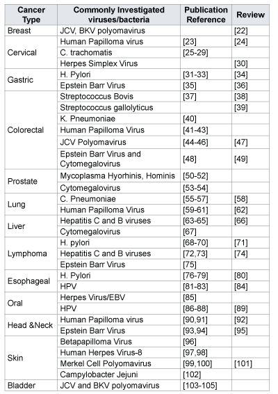
    Table 1: Cancer types and commonly investigated viruses and bacteria with selected references.

    Discussion

    From the portfolio analysis of NCI-funded grants some major themes were observed. These themes trend toward the characterization of overall microbiota community structure over a disease’s time course and in relation to various exposure events. Similarly, currently research on microbiome in cancer epidemiology is being conducted to identify biomarkers of risk, progression, and prognosis of cancer. Assessing the role of select microbiota or relative composition of microbiota, in disease prevention or progression is also a current trend.

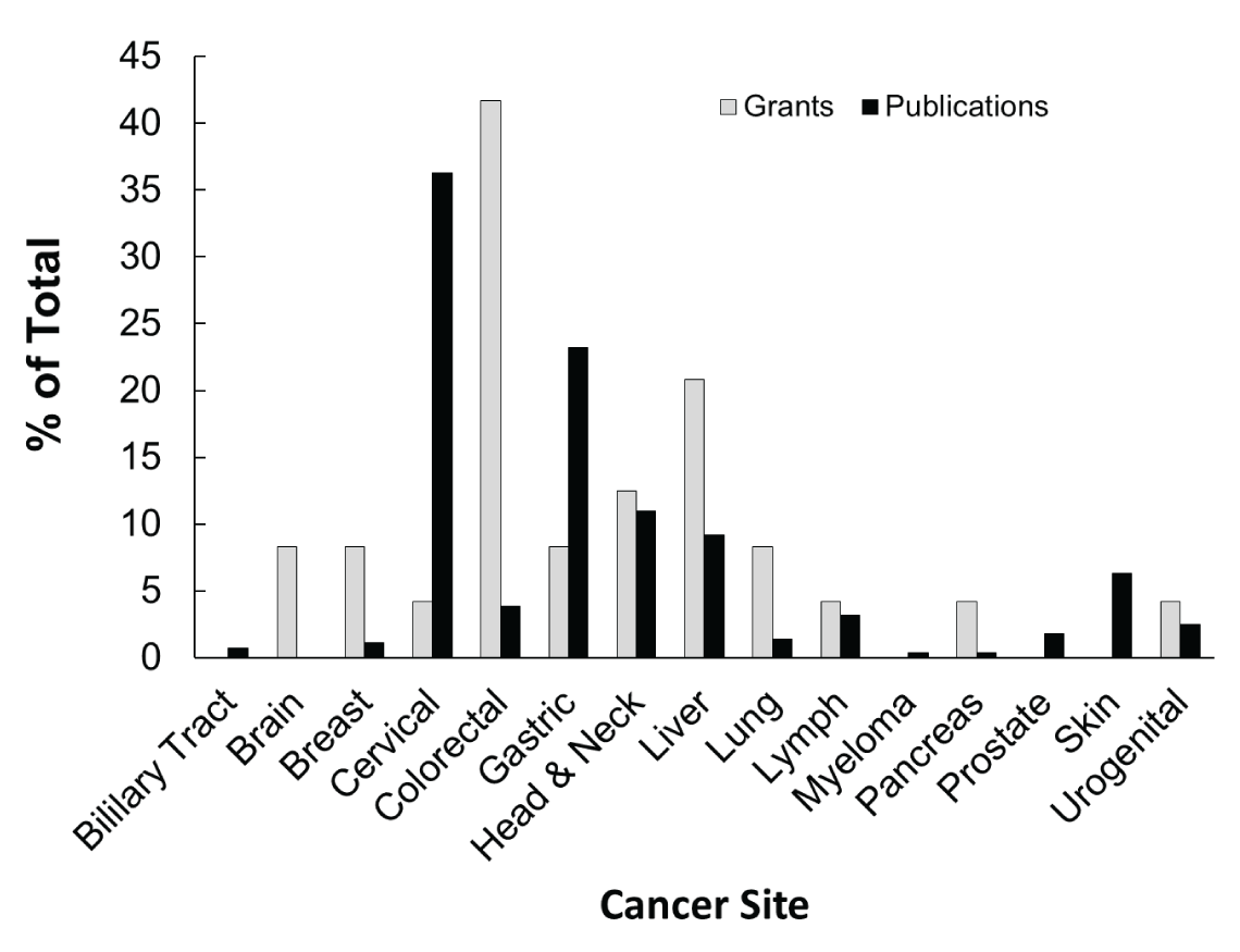
    Figure 1: Percentage of the total number of NCI-supported grants or PubMed publications, categorized by cancer site, that have incorporated microbiome analysis in cancer epidemiology.

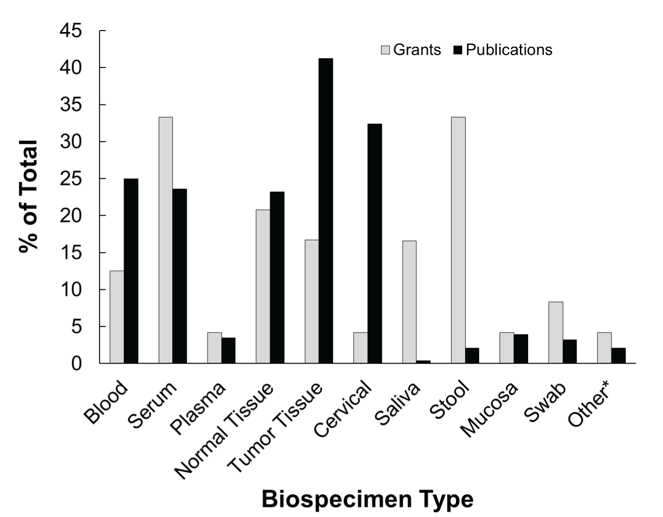
    Figure 2: Most commonly collected biospecimens types for microbiome analysis in cancer epidemiology. Percentage of the total number of NCIsupported grants or PubMed publications that have utilized the different biospecimens for microbiome analysis are shown. *Other includes: Eyebrow Hair, Oral Rinse/Wash, Bile, Nasal Mucus, and Urine.

    The majority of publications that were included in this analysis investigated known carcinogenic infectious agents such as HPV and cervical cancer, rather than what some might consider traditional microbiome studies. The reason for the inclusion of these studies in our analysis is in keeping with the definition of ‘microbiota’ to include viruses, which traditionally have been more commonly associated with cancer. This taken together, with the inclusion criteria of at least 100 cases, resulted in a limited number of true microbiome studies within the published literature. This demonstrates a gap in the research and also a possible starting point for inclusion of microbiome analysis in established cohorts or cohorts with existing samples. It is also recommended that cancer grants that proposed microbiome analysis and investigating novel associations in less studied cancers, such as pancreatic cancer and ovarian cancer, be keenly considered for funding.

    From this analysis, some challenges and knowledge gaps have been identified and outlined in table 1. In order to successfully analyze the microbiome over time in relation to pre-cancerous states, evidence supporting the stability of various sample types over time and at various temperatures is needed in order to establish guidelines for all epidemiologic studies of the microbiome. This has been done for stool samples [20] but confirmation is needed for the various other biospecimen types [21].

    Standardization of methods for reliable data integration across platforms and research centers is critical moving forwards. A large amount of sequence data already exists from microbiome research; however, this data was obtained using several different platforms and analysis softwares or pipelines available, each having inherent biases creating inaccurate data. Thus, it is critical to identify the biases for each and develop software that can correct and harmonize existing data for further research analysis. Standardization of methods, along with improvements to the reliability of available technology, will be paramount to streamlining the use of microbiome analysis in cancer epidemiology

    Though much of the work with the gut microbiome and colorectal cancer has elucidated the importance of these interactions in promoting disease, a similar level of understanding for other cancers and microbiomes has yet to be achieved. In addition, the exact mechanisms occurring between the host and microbiota to influence and determine cancer progression is not yet understood for the majority of cancers as it has been for colorectal cancer. Similarly, we do not have information on potential effects resulting from interactions of the microbiota with other carcinogenic infectious agents and/or chemical agents, and how these interactions, and the resulting metabolic changes modify or determine disease progression.

    Regarding the potential harm or protection against carcinogenesis that might be the effect of the microbiome, one example is that of cancerassociated infectious agents. Chronic infections caused by the hepatitis B and C viruses, human papillomaviruses (HPV), and Helicobacter pylori (H. pylori) are reported to be responsible for approximately 15% of all human cancers. Interestingly, although many of the infectious agents that have been associated with cancer such as HPV, Epstein-Barr virus (EBV), and H. pylori are highly prevalent in the world, most infected individuals do not develop cancer but remain lifelong carriers. Malignancies associated with infectious agents may result from prolonged latency as a result of chronic infections.

    The microbiome is a resource that holds promise for cancer epidemiology; however, in order to reliably utilize the microbiome for all its potential, work must be done at the level of basic science research to better understand the disease promoting or protective mechanisms that are at play between the host and microbiota.

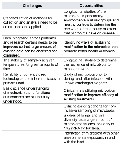
    Table 2: Challenges and opportunities identified for further research.

    Figure 3: Methods and technologies that are routinely being used for microbiome analysis in cancer epidemiology: The chart represents the percentage of the total number of NCI-supported grants or PubMed publications that are using the different methods and technologies. *Chromatography includes: liquid chromatography, gas chromatography, and chromatography combined with mass spectrometry. **Other includes: Giemsa Stain, histology, and clinical diagnostic tests.

    Presently, it is feasible to collect noninvasive samples representative of microbiomes from various potential cancer sites over multiple time points. This practice can easily be included in epidemiologic studies, which potentially may provide information on the natural history and disease progression, identification of those at highest risk, and ultimately inform research into specific mechanisms that lead to pre-cancerous states. Studying the microbiome in this way may also provide valuable information on how variables like diet and stress contribute to cancer risk.

    Opportunities for further study are presented in Table 1, which shows the commonly investigated bacteria or viruses and their associated cancer sites, may offer general starting points for investigation. Each of these cancer sites has an associated microbiome that can be sampled over time with the potential to yield information related to the natural history of a particular cancer. As a possible example of this, one might observe community shifts as a result of infection and ultimately progression to pre-cancer or cancer, allowing further risk stratification within a given population. Potential findings from longitudinal studies of the microbiomes of at risk populations may lead to the identification of better biomarkers for progression, which may allow for the implementation of more targeted and effective prevention strategies.

    In conclusion, the field of microbiome analysis offers a unique opportunity for cancer epidemiology which is beginning to receive a lot attention from the scientific community. If the challenges outlined in Table 2 are addressed, the microbiome, when combined with clinical and molecular epidemiology, may identify surrogate markers of disease progression for microbe-associated cancers, and may help in better cancer diagnosis, prevention, prognosis, and treatment outcomes assessment.

    Acknowledgements

    We acknowledge Dr. Jessica Pierce Ph.D, Genetics and Biochemistry Branch, NIDDK, NIH for reviewing early drafts of this manuscript.

    References

    1. Human Microbiome Project Consortium (2012) Structure, function and diversity of the healthy human microbiome. Nature 486: 207-214.[Ref.]
    2. Dominguez-Bello MG, Costello EK, Contreras M, Magris M, Hidalgo G, et al. (2010) Delivery mode shapes the acquisition and structure of the initial microbiota across multiple body habitats in newborns. Proceedings of the National Academy of Sciences of the United States of America 107: 11971-11975.[Ref.]
    3. Jarchum I, Pamer EG (2011) Regulation of innate and adaptive immunity by the commensal microbiota. Curr Opin Immunol 23: 353- 360.[Ref.]
    4. Martinez I, Muller CE, Walter J (2013) Long-term temporal analysis of the human fecal microbiota revealed a stable core of dominant bacterial species. PLoS One 8: e69621.[Ref.]
    5. Claesson MJ, Cusack S, O’Sullivan O, Greene-Diniz R, de Weerd H, et al. (2011) Composition, variability, and temporal stability of the intestinal microbiota of the elderly. Proc Natl Acad Sci USA 108: 4586-4591.[Ref.]
    6. Turner ND, Ritchie LE, Bresalier RS, Chapkin RS (2013) The microbiome and colorectal neoplasia: environmental modifiers of dysbiosis. Curr Gastroenterol Rep 15: 346.[Ref.]
    7. Candela M, Guidotti M, Fabbri A, Brigidi P, Franceschi C, et al. (2011) Human intestinal microbiota: cross-talk with the host and its potential role in colorectal cancer. Crit Rev Microbiol 37: 1-14.[Ref.]
    8. Arthur JC, Jobin C (2013) The complex interplay between inflammation, the microbiota and colorectal cancer. Gut Microbes 4: 253-258.[Ref.]
    9. Weir TL, Manter DK, Sheflin AM, Barnett BA, Heuberger AL, et al. (2013) Stool microbiome and metabolome differences between colorectal cancer patients and healthy adults. PLoS One 8: e70803.[Ref.]
    10. Ou J, Carbonero F, Zoetendal EG, DeLany JP, Wang M, et al. (2013) Diet, microbiota, and microbial metabolites in colon cancer risk in rural Africans and African Americans. Am J Clin Nutr 98: 111-120.[Ref.]
    11. Ou J, DeLany JP, Zhang M, Sharma S, O’Keefe SJ (2012) Association between low colonic short-chain fatty acids and high bile acids in high colon cancer risk populations. Nutr Cancer 64: 34-40.[Ref.]
    12. Wu N, Yang X, Zhang R, Li J, Xiao X, et al. (2013) Dysbiosis signature of fecal microbiota in colorectal cancer patients. Microb Ecol 66: 462- 470.[Ref.]
    13. Chen HM, Yu YN, Wang JL, Lin YW, Kong X, et al. (2013) Decreased dietary fiber intake and structural alteration of gut microbiota in patients with advanced colorectal adenoma. Am J Clin Nutr 97: 1044-1052.[Ref.]
    14. Amirian ES, Petrosino JF, Ajami NJ, Liu Y, Mims MP, et al. (2013) Potential role of gastrointestinal microbiota composition in prostate cancer risk. Infect Agent Cancer 8: 42.[Ref.]
    15. Michaud DS, Izard J (2014) Microbiota, oral microbiome, and pancreatic cancer. Cancer J 20: 203-206.[Ref.]
    16. Zambirinis CP, Pushalkar S, Saxena D, Miller G (2014) Pancreatic cancer, inflammation, and microbiome. Cancer J 20: 195-202.[Ref.]
    17. Farrell JJ, Zhang L, Zhou H, Chia D, Elashoff D, et al. (2012) Variations of oral microbiota are associated with pancreatic diseases including pancreatic cancer. Gut 61: 582-588.[Ref.]
    18. Schmidt BL, Kuczynski J, Bhattacharya A, Huey B, Corby PM, et al. (2014) Changes in abundance of oral microbiota associated with oral cancer. PLoS One 9: e98741.[Ref.]
    19. Henrich B, Rumming M, Sczyrba A, Velleuer E, Dietrich R, et al. (2014) Mycoplasma salivarium as a dominant coloniser of fanconi anaemia associated oral carcinoma. PLoS One 9: e92297.[Ref.]
    20. Carroll IM, Ringel-Kulka T, Siddle JP, Klaenhammer TR, Ringel Y (2012) Characterization of the fecal microbiota using high-throughput sequencing reveals a stable microbial community during storage. PLoS One 7: e46953.[Ref.]
    21. Aagaard K, Petrosino J, Keitel W, Watson M, Katancik J, et al. (2013) The Human Microbiome Project strategy for comprehensive sampling of the human microbiome and why it matters. FASEB J 27: 1012-1022.[Ref.]
    22. Alibek K, Kakpenova A, Mussabekova A, Sypabekova M, Karatayeva N (2013) Role of viruses in the development of breast cancer. Infect Agent Cancer 8: 32.[Ref.]
    23. Muñoz N, Bosch FX, De Sanjosé S, Herrero R, Castellsagué X, et al. (2003) Epidemiologic classification of human papillomavirus types associated with cervical cancer. N Engl J Med 348: 518-527.[Ref.]
    24. Steenbergen RDM, Snijders PJF, Heideman DAM, Meijer CJLM (2014) Clinical implications of (epi)genetic changes in HPV-induced cervical precancerous lesions. Nature Reviews Cancer 14: 395-405.[Ref.]
    25. Koskela P, Anttila T, Bjørge T, Brunsvig A, Dillner J, et al. (2000) Chlamydia trachomatis infection as a risk factor for invasive cervical cancer. Int J Cancer 85: 35-39.[Ref.]
    26. Anttila T, Saikku P, Koskela P, Bloigu A, Dillner J, et al. (2001) Serotypes of Chlamydia trachomatis and risk for development of cervical squamous cell carcinoma. JAMA 285: 47-51.[Ref.]
    27. Wallin KL, Wiklund F, Luostarinen T, Angstrom T, Anttila T, et al. (2002) A population-based prospective study of Chlamydia trachomatis infection and cervical carcinoma. Int J Cancer 101: 371-374.[Ref.]
    28. Safaeian M, Quint K, Schiffman M, Rodriguez AC, Wacholder S, et al. (2010) Chlamydia trachomatis and risk of prevalent and incident cervical premalignancy in a population-based cohort. J Natl Cancer Inst 102: 1794-1804.[Ref.]
    29. Smith JS, Bosetti C, Muñoz N, Herrero R, Bosch FX, et al. (2004) Chlamydia trachomatis and invasive cervical cancer: A pooled analysis of the IARC multicentric case-control study. Int J Cancer 111: 431-439.[Ref.]
    30. Cao S, Gan Y, Dong X, Lu Z (2014) Herpes simplex virus type 2 and the risk of cervical cancer: a meta-analysis of observational studies. Arch Gynecol Obstet 290: 1059-1066.[Ref.]
    31. Uemura N, Okamoto S, Yamamoto S, Matsumura N, Yamaguchi S, et al. (2001) Helicobacter pylori infection and the development of gastric cancer. N Engl J Med 345:784-789.[Ref.]
    32. Polk DB, Peek RM, Jr (2010) Helicobacter pylori: gastric cancer and beyond. Nat Rev Cancer 10: 403-414.[Ref.]
    33. Ma JL, Zhang L, Brown LM, Li JY, Shen L, et al. (2012) Fifteen-year effects of Helicobacter pylori, garlic, and vitamin treatments on gastric cancer incidence and mortality. J Natl Cancer Inst 104: 488-492.[Ref.]
    34. Brawner KM, Morrow CD, Smith PD (2014) Gastric microbiome and gastric cancer. Cancer J 20: 211-216.[Ref.]
    35. Osato T, Imai S (1996) Epstein-Barr virus and gastric carcinoma. Seminars in Cancer Biology 7: 175-182.[Ref.]
    36. Iizasa H, Nanbo A, Nishikawa J, Jinushi M, Yoshiyama H (2012) Epstein-Barr Virus (EBV)-associated gastric carcinoma. Viruses 4: 3420-3439.[Ref.]
    37. Ellmerich S, Schöller M, Duranton B, Gossé F, Galluser M, et al. (2000) Promotion of intestinal carcinogenesis by Streptococcus bovis. Carcinogenesis 21: 753-756.[Ref.]
    38. Abdulamir AS, Hafidh RR, Bakar FA (2011) The association of Streptococcus bovis/gallolyticus with colorectal tumors: The nature and the underlying mechanisms of its etiological role. J Exp Clin Cancer Res 30: 11.[Ref.]
    39. Boleij A, van Gelder MM, Swinkels DW, Tjalsma H (2011) Clinical Importance of Streptococcus gallolyticus infection among colorectal cancer patients: systematic review and meta-analysis. Clin Infect Dis 53: 870-878.[Ref.]
    40. Huang WK, Chang JW, See LC, Tu HT, Chen JS, et al. (2012) Higher rate of colorectal cancer among patients with pyogenic liver abscess with Klebsiella pneumoniae than those without: an 11-year follow-up study. Colorectal Dis 14: e794-e801.[Ref.]
    41. Lee YM, Leu SY, Chiang H, Fung CP, Liu WT (2001) Human papillomavirus type 18 in colorectal cancer. J Microbiol Immunol Infect 34: 87-91.[Ref.]
    42. Bodaghi S, Yamanegi K, Xiao SY, Da Costa M, Palefsky JM, et al. (2005) Colorectal papillomavirus infection in patients with colorectal cancer. Clin Cancer Res 11: 2862-2867.[Ref.]
    43. Chen TH, Huang CC, Yeh KT, Chang SH, Chang SW, et al. (2012) Human papilloma virus 16 E6 oncoprotein associated with p53 inactivation in colorectal cancer. World J Gastroenterol 18: 4051-4058.[Ref.]
    44. Laghi L, Randolph AE, Chauhan DP, Marra G, Major EO, et al. (1999) JC virus DNA is present in the mucosa of the human colon and in colorectal cancers. Proc Natl Acad Sci USA 96: 7484-7489.[Ref.]
    45. Newcomb PA, Bush AC, Stoner GL, Lampe JW, Potter JD, et al. (2004) No evidence of an association of JC virus and colon neoplasia. Cancer Epidemiol Biomarkers Prev 13: 662-666.[Ref.]
    46. Butcher LD, Garcia M, Arnold M, Ueno H, Goel A, et al. (2014) Immune response to JC virus T antigen in patients with and without colorectal neoplasia. Gut microbes 5: 468-475.[Ref.]
    47. Niv Y, Goel A, Boland CR (2005) JC virus and colorectal cancer: A possible trigger in the chromosomal instability pathway. Curr Opin Gastroenterol 21: 85-89.[Ref.]
    48. Militello V, Trevisan M, Squarzon L, Biasolo MA, Rugge M, et al. (2009) Investigation on the presence of polyomavirus, herpesvirus, and papillomavirus sequences in colorectal neoplasms and their association with cancer. Int J Cancer 124: 2501-2503.[Ref.]
    49. Hasan N, Pollack A, Cho I (2010) Infectious Causes of Colorectal Cancer. Infect Dis Clin North Am 24: 1019-1039.[Ref.]
    50. Namiki K, Goodison S, Porvasnik S, Allan RW, Iczkowski KA, et al. (2009) Persistent exposure to mycoplasma induces malignant transformation of human prostate cells. PLoS One 4: e6872.[Ref.]
    51. Urbanek C, Goodison S, Chang M, Porvasnik S, Sakamoto N, et al. (2011) Detection of antibodies directed at M. hyorhinis p37 in the serum of men with newly diagnosed prostate cancer. BMC cancer 11: 233.[Ref.]
    52. Barykova YA, Logunov DY, Shmarov MM, Vinarov AZ, Fiev DN, et al. (2011) Association of Mycoplasma hominis infection with prostate cancer. Oncotarget 2: 289-297.[Ref.]
    53. Samanta M, Harkins L, Klemm K, Britt WJ, Cobbs CS (2003) High prevalence of human cytomegalovirus in prostatic intraepithelial neoplasia and prostatic carcinoma. J Urol 170: 998-1002.[Ref.]
    54. Sutcliffe S, Till C, Gaydos CA, Jenkins FJ, Goodman PJ, et al. (2012) Prospective study of cytomegalovirus serostatus and prostate cancer risk in the Prostate Cancer Prevention Trial. Cancer Causes Control 23: 1511-1518.[Ref.]
    55. Laurila AL, Anttila T, Läärä E, Bloigu A, Virtamo J, et al. (1997) Serological evidence of an association between Chlamydia pneumoniae infection and lung cancer. Int J Cancer 74: 31-34.[Ref.]
    56. Littman AJ, Jackson LA, Vaughan TL (2005) Chlamydia pneumoniae and lung cancer: Epidemiologic evidence. Cancer Epidemiol Biomarkers Prev 14: 773-778.[Ref.]
    57. Chaturvedi AK, Gaydos CA, Agreda P, Holden JP, Chatterjee N, et al. (2010) Chlamydia pneumoniae infection and risk for lung cancer. Cancer Epidemiol Biomarkers Prev 19: 1498-1505.[Ref.]
    58. Zhan P, Suo LJ, Qian Q, Shen XK, Qiu LX, et al. (2011) Chlamydia pneumoniae infection and lung cancer risk: A meta-analysis. Eur J Cancer 47: 742-747.[Ref.]
    59. Yousem SA, Ohori NP, Sonmez-Alpan E (1992) Occurrence of human papillomavirus DNA in primary lung neoplasms. Cancer 69: 693-697[Ref.]
    60. Klein F, Amin Kotb WF, Petersen I (2009) Incidence of human papilloma virus in lung cancer. Lung Cancer. 65:13-18.[Ref.]
    61. Galvan A, Noci S, Taverna F, Lombardo C, Franceschi S, et al. (2012) Testing of human papillomavirus in lung cancer and non-tumor lung tissue. BMC cancer 12: 512.[Ref.]
    62. Syrjänen KJ (2002) HPV infections and lung cancer. J Clin Pathol 55: 885-891.[Ref.]
    63. Saito I, Miyamura T, Ohbayashi A, Harada H, Katayama T, et al. (1990) Hepatitis C virus infection is associated with the development of hepatocellular carcinoma. Proc Natl Acad Sci USA 87: 6547-6549.[Ref.]
    64. Chen CJ, Yang HI, Su J, Jen CL, You SL, et al. (2006) Risk of hepatocellular carcinoma across a biological gradient of serum hepatitis B virus DNA Level. JAMA 295: 65-73.[Ref.]
    65. Perz JF, Armstrong GL, Farrington LA, Hutin YJF, Bell BP (2006) The contributions of hepatitis B virus and hepatitis C virus infections to cirrhosis and primary liver cancer worldwide. J Hepatol 45: 529-538.[Ref.]
    66. Arzumanyan A, Reis HMGPV, Feitelson MA (2013) Pathogenic mechanisms in HBV- and HCV-associated hepatocellular carcinoma. Nature Reviews Cancer 13: 123-135.[Ref.]
    67. Lepiller Q, Tripathy MK, Di Martino V, Kantelip B, Herbein G (2011) Increased HCMV seroprevalence in patients with hepatocellular carcinoma. Virol J 8:485.[Ref.]
    68. Parsonnet J, Hansen S, Rodriguez L, Gelb AB, Warnke RA, et al. (1994) Helicobacter pylori infection and gastric lymphoma. N Engl J Med 330: 1267-1271.[Ref.]
    69. Choi YJ, Kim N, Paik JH, Kim JM, Lee SH, et al. (2013) Characteristics of Helicobacter pylori-positive and Helicobacter pylori-negative gastric mucosa-associated lymphoid tissue lymphoma and their influence on clinical outcome. Helicobacter 18: 197-205.[Ref.]
    70. Pereira MI, Medeiros JA (2014) Role of Helicobacter pylori in gastric mucosa-associated lymphoid tissue lymphomas. World J Gastroenterol 20: 684-698.[Ref.]
    71. Zullo A, Hassan C, Ridola L, Repici A, Manta R, et al. (2014) Gastric MALT lymphoma: old and new insights. Ann Gastroenterol 27: 27-33.[Ref.]
    72. Mizorogi F, Hiramoto J, Nozato A, Takekuma Y, Nagayama K, et al. (2000) Hepatitis C virus infection in patients with B-cell non-Hodgkin’s lymphoma. Intern Med 39: 112-117.[Ref.]
    73. Engels EA, Cho ER, Jee SH (2010) Hepatitis B virus infection and risk of non-Hodgkin lymphoma in South Korea: A cohort study. Lancet Oncol 11: 827-834.[Ref.]
    74. Dal Maso L, Franceschi S (2006) Hepatitis C virus and risk of lymphoma and other lymphoid neoplasms: A meta-analysis of epidemiologic studies. Cancer Epidemiol Biomarkers Prev 15: 2078-2085.[Ref.]
    75. Zhang Y, Peng J, Tang Y, He J, Peng J, et al. (2010) The prevalence of Epstein-Barr virus infection in different types and sites of lymphomas. Jpn J Infect Dis 63: 132-135.[Ref.]
    76. Chow WH, Blaser MJ, Blot WJ, Gammon MD, Vaughan TL, et al. (1998) An inverse relation between cagA+ strains of Helicobacter pylori infection and risk of esophageal and gastric cardia adenocarcinoma. Cancer Res 58: 588-590.[Ref.]
    77. Wu AH, Crabtree JE, Bernstein L, Hawtin P, Cockburn M, et al. (2003) Role of Helicobacter pylori CagA+ strains and risk of adenocarcinoma of the stomach and esophagus. Int J Cancer 103: 815-821.[Ref.]
    78. Cook MB, Dawsey SM, Diaw L, Blaser MJ, Perez-Perez GI, et al. (2010) Serum pepsinogens and Helicobacter pylori in relation to the risk of esophageal squamous cell carcinoma in the alpha-tocopherol, beta-carotene cancer prevention study. Cancer Epidemiol Biomarkers Prev 19: 1966-1975.[Ref.]
    79. Thrift AP, Pandeya N, Smith KJ, Green AC, Hayward NK, et al. (2012) Helicobacter pylori infection and the risks of Barrett’s oesophagus: a population-based case-control study. Int J Cancer 130: 2407-2416.[Ref.]
    80. Peek Jr RM, Blaser MJ (2002) Helicobacter pylori and gastrointestinal tract adenocarcinomas. Nat Rev Cancer 2: 28-37.[Ref.]
    81. De Villiersi EM, Gunst K, Stein H, Scherübl H (2004) Esophageal squamous cell cancer in patients with head and neck cancer: Prevalence of human papillomavirus DNA sequences. Int J Cancer 109: 253-258.[Ref.]
    82. Liyanage SS, Segelov E, Garland SM, Tabrizi SN, Seale H, et al. (2013) Role of human papillomaviruses in esophageal squamous cell carcinoma. Asia-Pacific journal of clinical oncology 9: 12-28.[Ref.]
    83. Goto A, Li CP, Ota S, Niki T, Ohtsuki Y, et al. (2011) Human papillomavirus infection in lung and esophageal cancers: analysis of 485 Asian cases. J Med Virol 83: 1383-1390.[Ref.]
    84. Syrjänen K (2010) Human papillomavirus (HPV) involvement in esophageal carcinogenesis. International Journal of Cancer Research and Prevention 3: 205-242.
    85. Jalouli J, Jalouli MM, Sapkota D, Ibrahim SO, Larsson PA, et al. (2012) Human papilloma virus, herpes simplex virus and epstein barr virus in oral squamous cell carcinoma from eight different countries. Anticancer Res 32: 571-580.[Ref.]
    86. Smith EM, Ritche JM, Summersgill KF, Hoffman HT, Wang DH, et al. (2004) Human papillomavirus in oral exfoliated cells and risk of head and neck cancer. J Natl Cancer Inst 96: 449-455.[Ref.]
    87. Ragin CCR, Modugno F, Gollin SM (2007) The epidemiology and risk factors of head and neck cancer: A focus on human papillomavirus. J Dent Res 86: 104-114.[Ref.]
    88. Chaturvedi AK, Engels EA, Pfeiffer RM, Hernandez BY, Xiao W, et al. (2011) Human papillomavirus and rising oropharyngeal cancer incidence in the United States. J Clin Oncol 29: 4294-4301.[Ref.]
    89. Hu FL, Yu YC (2013) Progress on role of human papilloma virus (HPV) in oropharyngeal cancer. Fudan University Journal of Medical Sciences 40: 482-485.
    90. Hu FL, Yu YC (2013) Progress on role of human papilloma virus (HPV) in oropharyngeal cancer. Fudan University Journal of Medical Sciences 40: 482-485.[Ref.]
    91. Dayyani F, Etzel CJ, Liu M, Ho CH, Lippman SM, et al. (2010) Metaanalysis of the impact of human papillomavirus (HPV) on cancer risk and overall survival in head and neck squamous cell carcinomas (HNSCC). Head Neck Oncol 2: 15.[Ref.]
    92. Syrjänen S (2005) Human papillomavirus (HPV) in head and neck cancer. J Clin Virol 32: S59-S66.[Ref.]
    93. Chan KH, Gu YL, Ng F, Ng PS, Seto WH, et al. (2003) EBV specific antibody-based and DNA-based assays in serologic diagnosis of nasopharyngeal carcinoma. Int J Cancer 105: 706-709.[Ref.]
    94. Wang WY, Twu CW, Chen HH, Jan JS, Jiang RS, et al. (2010) Plasma EBV DNA clearance rate as a novel prognostic marker for metastatic/ recurrent nasopharyngeal carcinoma. Clin Cancer Res 16: 1016-1024.[Ref.]
    95. Goldenberg D, Golz A, Netzer A, Rosenblatt E, Rachmiel A, et al. (2001) Epstein-Barr virus and cancers of the head and neck. American journal of otolaryngology 22: 197-205.[Ref.]
    96. Bouwes Bavinck JN, Neale RE, Abeni D, Euvrard S, Green AC, et al. (2010) Multicenter study of the association between betapapillomavirus infection and cutaneous squamous cell carcinoma. Cancer Res 70: 9777-9786.[Ref.]
    97. Patel RM, Goldblum JR, Hsi ED (2004) Immunohistochemical detection of human herpes virus-8 latent nuclear antigen-1 is useful in the diagnosis of Kaposi sarcoma. Mod Pathol 17: 456-460.[Ref.]
    98. Pak F, Pyakural P, Kokhaei P, Kaaya E, Pourfathollah AA, et al. (2005) HHV-8/KSHV during the development of Kaposi’s sarcoma: Evaluation by polymerase chain reaction and immunohistochemistry. Journal J Cutan Pathol 32: 21-27.[Ref.]
    99. Kassem A, Technau K, Kurz AK, Pantulu D, Löning M, et al. (2009) Merkel cell polyomavirus sequences are frequently detected in nonmelanoma skin cancer of immunosuppressed patients. Int J Cancer 125: 356-361.[Ref.]
    100. Rollison DE, Giuliano AR, Messina JL, Fenske NA, Cherpelis BS, et al. (2012) Case-control study of Merkel cell polyomavirus infection and cutaneous squamous cell carcinoma. Cancer Epidemiol Biomarkers Prev 21:74-81.[Ref.]
    101. Gjoerup O, Chang Y (2010) Update on human polyomaviruses and cancer. Adv Cancer Res 106: 1-51.[Ref.]
    102. Brauner A, Brandt L, Frisan T, Thelestam M, Ekbom A (2010) Is there a risk of cancer development after Campylobacter infection? Scand J Gastroenterol 45: 893-897.[Ref.]
    103. . Fioriti D, Pietropaolo V, Dal Forno S, Laurenti C, Chiarini F, et al. (2003) Urothelial bladder carcinoma and viral infections: Different association with human polyomaviruses and papillomaviruses. Int J Immunopathol Pharmacol 16: 283-288.[Ref.]
    104. Robles C, Viscidi R, Malats N, Silverman DT, Tardon A, et al. (2013) Bladder cancer and seroreactivity to BK, JC and Merkel cell polyomaviruses: the Spanish bladder cancer study. Int J Cancer 133: 597-603.[Ref.]
    105. Panagiotakis GI, Papadogianni D, Chatziioannou MN, Lasithiotaki I, Delakas D, et al. (2013) Association of human herpes, papilloma and polyoma virus families with bladder cancer. Tumour Biol 34:71-79.[Ref.]

    Download Provisional pdf here