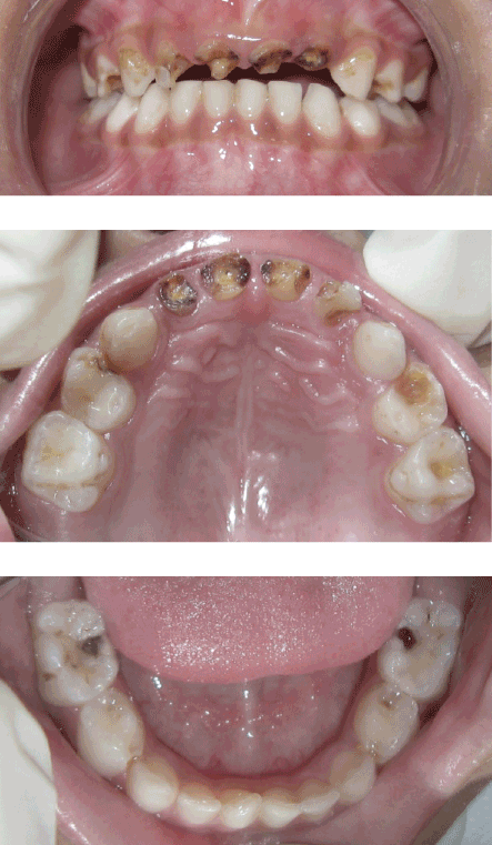
Figure 1: Preoperative view of child’s decayed incisors

Neeti Mittal1,* Praveen Kumar2
1Department of Pediatric and Preventive Dentistry, Santosh Dental College and Hospital, Ghaziabad, Uttar Pradesh, India*Corresponding author: Neeti Mittal, Department of Pediatric and Preventive Dentistry, Santosh Dental College and Hospital, No 1 Santosh Nagar, Pratap Vihar, Ghaziabad, Uttar Pradesh, India, E-mail: dr.neetipgi@gmail.com
Due to increasing concerns over self appearance, adequate consideration to esthetics in addition to functional aspects are necessary part of rehabilitation of mutilated teeth to help children grow into a psychologically balanced personality. The present article describes management of a pediatric patient with grossly broken down anterior teeth employing indirect composite restorations using a modified technique. This technique provided an excellent esthetic solution with less chair-side time.
Composite; Esthetics; Indirect; Mutilated teeth; Primary dentition
The responsibility of a pediatric dentist is not limited to mere replacement of lost tooth structure, but also the confidence of the young child. Giving the right magnitude of attention to esthetic appearance of a child’s dentition is necessary to prevent the present as well as future unpleasant psychological sequelae. The desire to have beautiful restorations is rightful privilege of our young clients. From here arose the need to look for various techniques which take into consideration the esthetic component while not compromising on functional aspects such as composite restorations with celluloid strip crowns [1], indirect resin composite crowns [2], stainless steel crowns with composite facing [3] and biologic restorations [4].
Although rehabilitation with various types of direct restorations mentioned above provide satisfactory esthetics; these can sometimes become problematic in a young child with behavioral issues. On the other hand, indirect restorations can still be performed owing to requirement for lesser chair-side time. Additional clinical benefits include precise marginal integrity [5], wear resistance [6], ideal proximal contacts [7], and excellent anatomic morphology [7].
The objective of this article is to present pediatric cases where rehabilitation of mutilated teeth was performed using indirect composite restorations.
A 4½ year-old boy, conscious of his appearance, presented to the Dental OPD of Jay Pee Hospital, Noida with the chief complaint of poor facial appearance due to discoloured and decayed front teeth. On examination, the child was found to have multiple carious lesions with root stumps of the maxillary primary central incisors (51,61,62) and grossly decayed 52 (Figure 1). Carious lesions were seen in 53, 54, 55, 63, 64, 65, 74, 75, 84, 85. Clinically, the root stumps of 51,61 and 62 were found to be firm, with an extension of the remaining crown approximately ≥ 1 mm above the gingival margin. An intraoral peri apical radiograph of these teeth showed intact roots and normal development of permanent successors. The management of this patient consisted of, giving complete preventive care, along with, restoration of all decayed teeth with direct as well as indirect composite restorations.

Figure 1: Preoperative view of child’s decayed incisors
Endodontic treatment and esthetic rehabilitation of grossly decayed maxillary incisors was carried out was carried out as mentioned herewith:
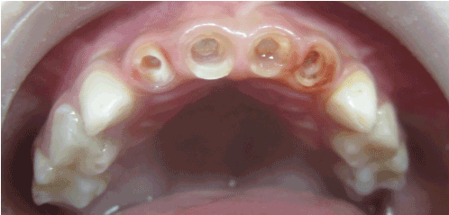
Figure 2: Final preparation
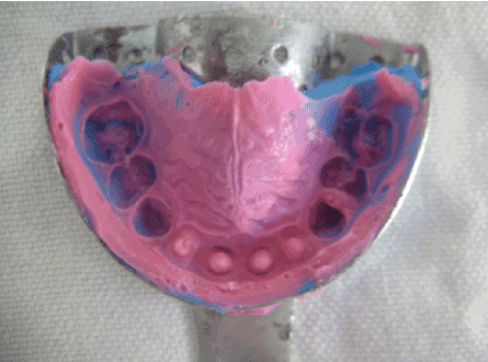
Figure 3: Final impression
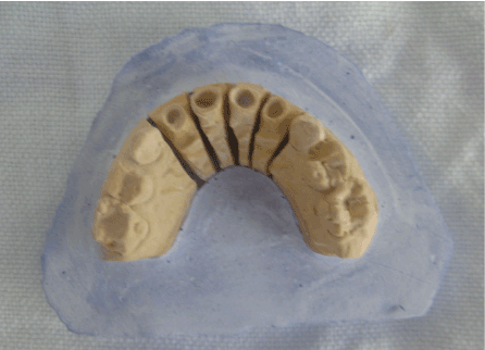
Figure 4a: Final cast after die cutting
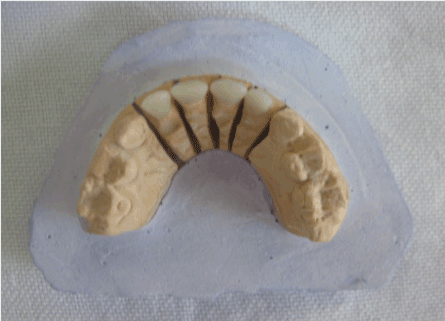
Figure 4b: Crowns seated on cast
This appointment was scheduled after a follow up period of one month to ensure success of endodontic treatment.
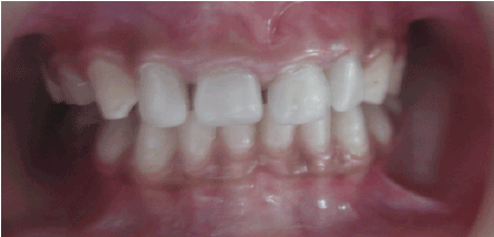
Figure 5: Postoperative view of restored maxillary incisors
The management of totally broken down primary teeth is carried out in two phases, core build up followed by restoration of the crown anatomy. A multitude of methods have been used for intra canal reinforcement for anterior teeth such as composite posts [1], short wire posts (omega loop) [8], Ni-Cr coil spring posts [2], readymade glass fibre posts [9], ribbond [10] and dentinal posts [11]. The crown anatomy can be restored by direct composite build up by incremental method [8], composite build up using celluloid strip crowns [1,9], composite build up by indirect technique [2,10], open faced stainless steel crowns [3], resin veneered stainless steel crowns [12], enamel veneers, and biological shell crowns [4,11].
Previously few authors have reported restoring grossly broken down anterior teeth by indirect technique using various types of posts such as preformed Ni-Cr posts [2], fiberglass posts and ribbond [10] as intra canal reinforcement. Instead of two step technique, in the present case reports composite crown and post were fabricated as a single unit by indirect method, thus saving the chair side time. All of the methods listed above require longer chair side time which may compromise the cooperation by young child with short attention span and little patience.
Although direct composite restorations form backbone of restorative dentistry, they have various limitations. These include post-operative pain due to contraction of the resin which is bonded to the thin cavity walls [13], marginal micro leakage following polymerization shrinkage especially at the cervical cavo surface margins [5], improper contact points [7] and relatively low wear resistance [6]. Extra-oral improved curing of the composite resin [7] can minimize above mentioned disadvantages of direct composite restorations.
An overabundance of systems for laboratory processed indirect composites could have tempted us to choose one for this case. Ignoring all the advantages of the commercially available indirect systems, i.e., greater filler loading for improved mechanical strength and better handling properties; we selected the composite material being routinely used for direct restorative procedures in our clinic. It was cured with same light cure unit being used for direct composite restorations. The rationale for this was to avoid greater economic burden owing to higher cost. In fact, these indirect composite systems were developed for permanent teeth which have a much longer time period to serve in oral cavity than primary teeth. For these reasons we preferred to use direct composite material.
Teeth lost in the anterior region infrequently require space maintenance, but there rehabilitation is important for speech and psychological well being of young children. Due to increasing concerns over esthetic appearance, metal crowns are no longer acceptable. It was seen that the restoration carried out with this technique offered a greater satisfaction to the patient, parent and the dentist himself.
With the present technique not only function was restored but also excellent esthetics was obtained with lesser chair side time. Thus, the technique presented here can help paediatric dentists enjoy the satisfaction of restoring the smiles of their young patrons with efficient handling of behavioral issues.
Download Provisional PDF Here
Article Type: Case Report
Citation: Mittal N, Kumar P (2015) Restoring the Young Smile via Indirect Composite Restorations. Int J Dent Oral Health 2(2): doi http://dx.doi. org/10.16966/2378-7090.163
Copyright: © 2015 Mittal N, et al. This is an open-access article distributed under the terms of the Creative Commons Attribution License, which permits unrestricted use, distribution, and reproduction in any medium, provided the original author and source are credited.
Publication history:
All Sci Forschen Journals are Open Access