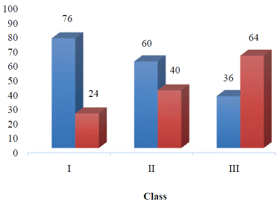
Figure 1: Results of Chi-Square test showing significant difference (p = 0.004) in ± 1 SD Bolton TSD distribution between Angle split groups in TMD group.

Markus Greven1,2* Eva Piehslinger2 Armin Mokhtari Tavana2,3
1Private Office, Bonn, Germany*Corresponding author: Markus Greven, Private Office, Bonn, Germany, Tel: 49 228 985900; E-mail: markusgreven@t-online.de
Objective: The aim of this retrospective study was to investigate the correlation between the Bolton Index (tooth size discrepancy TSD) and signs and symptoms of temporomandibular disorder (TMD).
Methods: One hundred fifty patients were divided into two groups: TMD group (75 patients diagnosed with an occlusion-dictated TMD according to the clinical protocol of the Vienna School of Interdisciplinary Dentistry (VieSID)) and a control group. Angle classification and Bolton Index (by using 3D Shape TRIOS intraoral scanner and 3Shape Ortho Analyzer software) were analyzed.
Results: Angle Class III patients showed a significantly greater prevalence of TSD compared to Class I and II in the TMD group. Forty-six out of the 150 patients were found with anterior TSD greater than ± 1 SD using Bolton Index.
Discussion: With the Bolton Index malocclusions can be measured and risk patients identified. The results of this study confirm the importance of a detailed diagnosis before orthodontic treatment.
Bolton Index; Tooth size discrepancy; Temporomandibular disorder; Angle classification
The temporomandibular joints (TMJ), the jaw muscles and/or the related structures are affecting by a heterogeneous group of conditions [1].
Rearrangement of the load-bearing joints is an important adaptive mechanism required for the proper distribution and operation of pressure. When the capacity of the rehabilitation joint is exceeded, the rehabilitation is associated with osteoarthritis [2,3] associated with “internal disc herniation of the TMJ” [4].
Wilkes CH [5] has developed a five-phase program to differentiate internal confusion based on clinical diagnostic and imaging principles (Table 1).
| Stage | Clinical features | Imaging |
| I. Early | Painless clicking; unrestricted function | Mild disc displacement; |
| Normal condyle | ||
| II. Early/intermediate | Intermittent painful clicking and locking | Mild disc displacement and |
| deformity; normal condyle | ||
| III. Intermediate | Frequent joint pain and locking; painful restricted function | Moderate disc displacement |
| and deformity; normal condyle | ||
| IV. Intermediate/late | Chronic pain and restricted mandibular function | Severe disc displacement and |
| deformity; abnormal condyle | ||
| V. Late gross | Severe joint dysfunction (crepitus) with variable pain | Severe disc displacement with perforation and deformity; |
| degenerative condylar changes |
Table 1: TMJ internal derangement classification of Wilkes [5].
Clinical observations demonstrate that numerous factors as trauma, unstable occlusion, functional overloading, Parafunctional and increased joint friction may affect the progression of TMD and associated degenerative changes [6,7].
A tooth size discrepancy (TSD) is defined as a disproportion among the sizes of individual teeth. When the maxillary and mandibular teeth are proportional in size a good occlusion with the correct overjet and overbite would be achieved. The best-known study of tooth size disharmony in relation to the treatment of malocclusion was by Bolton WA in 1958. Bolton developed ratios for estimating TSD by measuring the summed mesiodistal (MD) widths of the mandibular to the maxillary anterior teeth for orthodontic diagnosis. In another paper, Bolton expanded on the clinical use of dental size analysis. Bolton's general deviation from his original sample has been used to determine the need for dental reduction by internal removal or addition of dental tissue with restorative techniques [8].
By using digital software and tridimensional virtual models rapid and exact measurements are obtained [9,10].
Several studies have evaluated patients with different malocclusion groups (Class I, Class II, and Class III) that were orthodontically treated and found no statistically significant differences in the prevalence of TSD among the three groups [11-13]. However, other studies have observed significant differences in the presence of TSD among malocclusion groups [14-17].
The relationship between tooth decay and temporomandibular disorders (TMD) remains a controversial topic in dentistry. Indeed, while the orofacial pain experts seem to have adopted the “biopsychosocial model of TMD” [18], within the broader context of orofacial pain conditions [19], specialists focused on studying and restoring dental occlusion (i.e, orthodontists, prosthodontists, restorative dentists) have traditionally been declined the importance of occlusal dogmas [20].
Dental Occlusion is the main subject of Dentistry. Years of research have consistently illuminated many of the issues related to occlusion management in clinical practice [21]. A hypothetical relationship based on ‘malocclusion’ and TMD has been recommended for years by gnathology [22], but the occlusal paradigm of TMD have never been confirmed by faith [23]. Considering that the safe management of TMD symptoms is almost always enough to achieve positive results [24], and that chronic pain studies are people with a certain personality and not occlusal profiles [25-27]. After all, malocclusion can cause TMD and after a while, signs and symptoms of TMD appear [28- 31]; with the Bolton Index malocclusions can be measured. Based on these premises, the aim of this study is the evaluation the correlation between Bolton Index (tooth size discrepancy TSD) and signs and symptoms of temporomandibular disorders.
Accordingly, the hypothesis for this study is:
» There could be an association between Bolton Index and temporomandibular dysfunction signs and symptoms.
.A retrospective cohort study (with the control group) was done with 150 patients divided into two groups: the TMD group and a control group (normal population) with 75 patients each.
The selection criteria were:
» Males and females between the age of 18-28.
» No orthodontic treatments and no extraction (except 3.M).
» Previously obtained informed consent to use patient data.
All patients out of the TMD group took part in a standardized process according to the clinical protocol of the Vienna School of Interdisciplinary Dentistry (VieSID):
» Questionnaire and special medical analysis (recent infections, problems with cardiovascular, respiratory, digestive or metabolic system, allergies, urogenital problems, problems related to the central nervous system, psychological problems, rheumatic disease, hormonal disorders).
» Dental history analysis.
» Muscle and joint diagnosis (the state of muscles, both in the left and right sides of shoulders and neck, atlantooccipital regions, Posterior, medial and anterior temporalis muscles, deep and superficial masseter muscles, tuber maxillae muscles, pterygoideus medialis muscles, mylohyoideus muscles, digastricus muscles, supra and infra hyoidale muscles, laryngeal, sterno-cleido-mastoeideus, omohyoideus and tongue muscles; for bone and joint assessments, the examinations involved comparative palpation of jaw joints, lateral poles statically, lateral points in rotation, retral joint space and temporomandibular ligament).
» Occlusogram - Brux checker.
» X-ray- Panoramic, lateral cephalometric view.
The TMD subjects were diagnosed according to the appearance of objective/subjective symptoms and findings out of the above diagnostic measures.
Bolton Index measurement was done by 3D Shape TRIOS intraoral scanner and analysis by 3Shape Ortho Analyzer software (3Shape A/S, Copenhagen, Denmark).
With at least a 53% difference in the malfunction ratio between Angle Classes is predicted (effect size 0.43), at least 66 people, at least 22 persons in each class group at 90% power level, are required to work. Whether or not there is any difference in malfunctioning among the class groups will be investigated by the Chi-square test.
Data were collected and then analyzed with SPSS (version 21.) software. Demographic data were analyzed with descriptive tests and results were demonstrated by frequency (%) and Mean ± SD. Error analysis for all measurements, using the nonparametric Wilcoxon Statistical Test. For comparing groups and investigation of Bolton index and TMD signs and symptoms, the Chi-Square test was used. P-value is lesser than 0.05 was considered a significant correlation.
Finally, the patients were divided into groups by angle classification: TMD group (Class I, II (division 1,2), III) and Control group (Class I, II (division 1,2), III).
The mean age of the TMD group was 23.11 ± 3.19 years (with a range of 18-28 years), the mean age of the control group was 24.19 ± 2.78 years (with a range of 18-28 years).
The frequency of gender distribution within the studied groups is shown in table 2.
| Group | Male | Female | Total |
| TMD | 33 (44%) | 42 (56%) | 75 (100%) |
| Control | 36 (48%) | 39 (52%) | 75 (100%) |
| *Data were shown as frequency (percent). | |||
Table 2: Frequency of gender distribution in studied groups.
To gather data accurately, each tooth is measured in a large mesiodistal size using a digital caliper with an accuracy of 0.01 mm. Each dental size means that they were compared using ANOVA (Diversity Analysis) to determine whether dental size was related to gender, malocclusion classification, or both. No significant differences were found between the three groups where the size of each tooth was compared with the function of the Angle sections. Significant statistical difference (p> 0.05) was observed when each tooth size was compared to the sex function.
For the purpose of comparing the results of this study with those presented by Bolton WA [32], the data were categorized as “normal” for Bolton ratings within ± 1 SD (78.2 ± 1.55%) and “differences” in ratios greater than +1 SD.
A total of 42.7% of the TMD group and 18.7% of the control group in this study presented the Bolton TSD (greater than ± 1 SD). However, in both groups, no significant differences were observed between the three groups of malocclusion (p> 0.05) (Figures 1,2 and Tables 3,4).
Figure 1: Results of Chi-Square test showing significant difference (p = 0.004) in ± 1 SD Bolton TSD distribution between Angle split groups in TMD group.
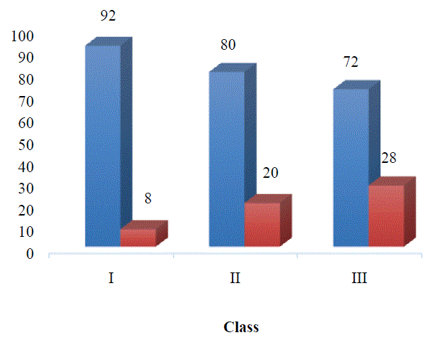
Figure 2: Results of Chi-Square test showing no significant difference (p = 0.217) in ± 1 SD Bolton TSD distribution between Angle split groups in the control group.
| Bolton Ratio | Class I | Class II | Class III | Total |
| Normal | 19 (76%) | 15 (60%) | 9 (36%) | 43 (57.4%) |
| Discrepancy | 6 (24%) | 10 (40%) | 16 (64%) | 32 (42.6%) |
| Total | 25 | 25 | 25 | 75 (100%) |
| *Data were shown as frequency (percent). | ||||
Table 3: Results of the Chi-Square test showing no significant difference (p = 0.004) in the spread of ± 1 SD Bolton TSD between Angle segmentation groups in the TMD group.
| Bolton Ratio | Class I | Class II | Class III | Total |
| Normal | 23 (92%) | 20 (80%) | 18 (72%) | 61 (81.4%) |
| Discrepancy | 2 (8%) | 5 (20%) | 7 (28%) | 14 (18.6%) |
| Total | 25 | 25 | 25 | 75 (100%) |
| *Data were shown as frequency (percent). | ||||
Table 4: Results of the Chi-Square test showing no significant difference (p = 0.217) in ± 1 SD Bolton TSD distribution between Angle segmentation groups in the control group.
Also, no significant differences were found in the spread of dental size in Bolton as a gender function in both groups (p> 0.05) (Figures 3,4 and Tables 5,6).
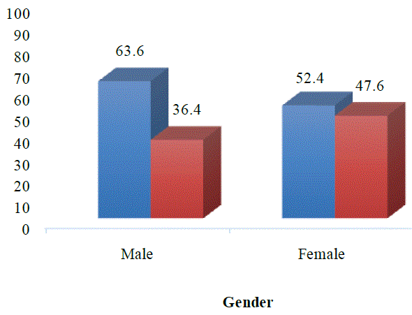
Figure 3: Chi-Square testing results show no significant difference (p = 0.387) in ± 1 SD Bolton TSD between sexes in the TMD group.
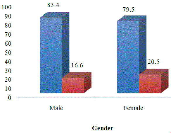
Figure 4: Results of Chi-Square test showing no significant difference (p = 0.277) in ± 1 SD Bolton TSD distribution between sexes in the control group.
| Anterior Ratio | Gender | Total | |
| Male | Female | ||
| Normal | 21 (63.6%) | 22 (52.4%) | 43 (57.4%) |
| Discrepancy | 12 (36.4%) | 20 (47.6%) | 32 (42.6%) |
| Total | 33 (44%) | 42 (56%) | 75 (100%) |
| *Data were shown as frequency (percent). | |||
Table 5: Chi-Square test results showing no significant difference (p = 0.387) in ± 1 SD Bolton TSD between sexes in the TMD group.
| Anterior Ratio | Gender | Total | |
| Male | Female | ||
| Normal | 30 (83.4%) | 31 (79.5%) | 61 (81.4%) |
| Discrepancy | 6 (16.6%) | 8 (20.5%) | 14 (18.6%) |
| Total | 36 (48%) | 39 (52%) | 75 (100%) |
| *Data were shown as frequency (percent). | |||
Table 6: Chi-Square test results not showing significant differences (p = 0.277) in spread ± 1 SD Bolton TSD between sexes in the control.
For analysis of the correlation of the Bolton Index with TMD signs and symptoms, a chi-square test was performed for each one. The results were shown in tables 7-9.
| Class I | Bolton Index | Total | ||
| Normal | Discrepancy | |||
| Sign | Normal | 19 (27.5%) | 0 |
19 (25.3%) |
| Abnormal | 3 (4.3%) | 3 (50%) | 6 (8%) | |
| Symptom | Normal | 22 (31.8%) | 0 |
22 (29.3%) |
| Abnormal | 2 (2.9%) | 1 (17.3%) | 3 (4.05%) | |
| Sign with symptom | Normal | 22 (31.8%) | 0 |
22 (29.3%) |
| Abnormal | 1 (1.7%) | 2 (32.7%) | 3 (4.05%) | |
| Total | 69 (92%) | 6 (8%) | 75 (100%) | |
Table 7: Chi-Square testing results show no significant difference (p = 0.098) in increasing symptoms and signs of ± 1 SD Bolton TSD and TMD symptoms within Class I.
| Class II | Bolton Index | Total | |||
| Normal | Discrepancy | ||||
| Sign | Normal | 15 (23%) | 0 | 15 (20%) | |
| Abnormal | 8 (12.3%) | 2 (20%) | 10 (13.3%) | ||
| Symptom | Normal | 20 (30.7%) | 0 | 20 (26.7) | |
| Abnormal | 2 (3%) | 3 (30%) | 5 (6.7%) | ||
| Sign with symptom | Normal | 19 (29.2%) | 0 | 19 (25.3%) | |
| Abnormal | 1 (1.8%) | 5 (50%) | 6 (8%) | ||
| Total | 65 (86.7%) | 10 (13.3%) | 75 (100%) | ||
Table 8: Chi-Square testing results not showing significant differences (p = 0.108) in increasing symptoms and signs of ± 1 SD Bolton TSD and TMD symptoms within Class II.
| Class III | Bolton Index | Total | |||
| Normal | Discrepancy | ||||
| Sign | Normal | 11 (18.6%) | 0 | 11 (14.7%) | |
| Abnormal | 8 (13.5%) | 6 (37.5%) | 14 (18.7%) | ||
| Symptom | Normal | 17 (28.8%) | 0 | 17 (22.7%) | |
| Abnormal | 5 (8.4%) | 3 (18.8%) | 8 (10.6%) | ||
| Sign with symptom | Normal | 16 (27.1%) | 0 | 16 (21.3%) | |
| Abnormal | 2 (3.6%) | 7 (43.7%) | 9 (12%) | ||
| Total | 59 (78.7%) | 16 (21.3%) | 75 (100%) | ||
Table 9: Chi-Square test results showing significant difference (p = 0.003) in increasing signs and symptoms of ± 1 SD Bolton TSD and TMD symptoms within Class III.
The main hypothesis for this study was that there is an association between Bolton Index and TMD signs and symptoms. After statistical analysis, no correlation has been found between TMD signs and symptoms in Class I (p = 0.098) and II (p = 0.108) with Bolton Index, in Class III there is a significant correlation (p = 0.003).
In the literature importance of TSD in orthodontic diagnosis has been reported. The prevalence of anterior TSD in this model is an indicator of how important it is to make a comprehensive diagnosis before orthodontic treatment. Forty-six individuals (31%) out of a total sample of 150 presented with an anterior TSD greater than ± 1 SD using the Bolton analysis parameter. This percentage (31%) found by Richardson ER and Malhotra SK [33] (33.7%) and Bolton WA [32] (29%) is significant.
In the case of TSD among Angle classifications, the results of this study shown a positive correlation; however, in the higher malocclusion class the rate of discrepancy was higher. The results of this study are consistent with the results of Nie Q and Lin J [17] after analyzing 360 Chinese people for TSD using Angle classification.
Dentists should be accustomed to differences in dental size in the early stages of diagnosis and treatment if success is achieved in orthodontic completion. TSD is considered an important variant, especially in the anterior segment.
Genetic effects are considered important in determining dental size, and early reports were related to clinical observations in families [34].
Gatorade emphasized genetic predisposition and described dental size determination as versatility, environment and nutrition play an important role [35].
With many other human characteristics, teeth vary in size between men and women. Gender differences have been reported in the literature and may be clinically relevant. According to Seipel, quoted by Lavelle CL [36], there are fewer gender differences in the main dental area than in permanent extraction. Male teeth are generally considered to be larger than female teeth [37,38]. In both basic and permanent implant teeth, the upper canines and upper incisors show the largest sex differences [38] while the upper lateral incisor and lower incisor are homogenous [37].
The first concerns raised in dental literature related to dental size date back to the 1920s. In various researches, the authors suggest that there should be a balance between upper and lower teeth [15,32,39]. Gilpatric and later Stanton studied 2000 people and found that the upper teeth should be 8 to 12 mm in size than the lower teeth and that a score of more than 8 to 12 mm can lead to overeating.
Several studies have been published that explain the importance of the correct proportion of tooth size between the upper and lower arches [40-42]. Lundstrom A [41] studied the relationship between mandibular and maxillary anterior sum and labeled it an “internal index”. With the right charge, the right rate is found to be from 73% to 85% that is 79%. Bolton WA [42] investigated 55 cases with a ‘very good’ closure. After analyzing dental sizes from canine to canine in the maxillary and mandibular arches, Bolton has developed an ideal internal balance of 77.2% with a standard deviation (SD) of 1.65%.
In the most recent articles, other variables such as incisor inclination [43,44], incisor size [45,46] and arch form have been described as important factors to consider in achieving good occlusion relationships. Efforts have been made to reconcile Bolton's analysis with diversity. Several authors [45-47] have proposed new ways to study TSD. However, these recommendations need to be tested in clinical studies and, in the meantime, Bolton’s analysis serves as an effective clinical tool for measuring the various interdependent dental interactions upwards.
A dentist who knows and is knowledgeable about a possible TSD will be better prepared to diagnose and plan treatment with more accurate certainty. These conclusions can have a significant impact on clinical decision-making and further studies should be done in this field.
The authors report no conflict of interest.
Download Provisional PDF Here
Article Type: RESEARCH ARTICLE
Citation: Greven M, Piehslinger E, Mokhtari Tavana A (2021) Correlation of Signs and Symptoms of Temporomandibular Disease with Bolton Index. Int J Dent Oral Health 7(5): dx.doi.org/10.16966/2378-7090.367
Copyright: © 2021 Greven M, et al. This is an open-access article distributed under the terms of the Creative Commons Attribution License, which permits unrestricted use, distribution, and reproduction in any medium, provided the original author and source are credited.
Publication history:
All Sci Forschen Journals are Open Access