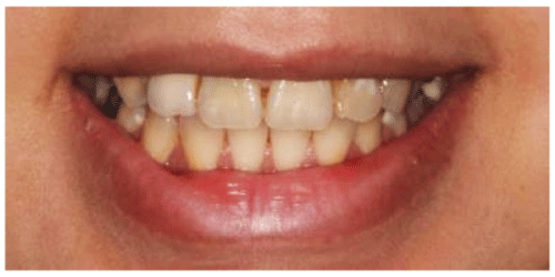
Figure 1: Pretreatment smile. The patient complained about dark yellow teeth, poor esthetics of the full coverage restoration in tooth #12, and black triangle between the central incisors.


Diemah F Alhekeir1* Rana A Alsarhan1 Abier F Alhekeir2
1Restorative Department, Ministry of Health, Riyadh, Saudi Arabia*Corresponding author: Diemah F Alhekeir, Restorative Department, Ministry of Health, 11176 Riyadh, Saudi Arabia, E-mail: d.alhekeir@gmail.com
Interaction among various dental specialists is essential for managing extensive dental work. Appropriate case management and dividing the required treatment into phases leads to successful treatment. In this case report, we describe a treatment sequence for managing full-mouth rehabilitation with complex prosthodontics. Phasing the treatment facilitated functional and aesthetic correction. Understanding of the treatment process and explaining the facts to the patient can avoid discrepancy in the outcomes.
Complex prosthodontics; Full-mouth rehabilitation; Case management; Multidisciplinary approach
Extensive evaluation, correct diagnosis, and development of a proper treatment plan is challenging when managing patients requiring full-mouth rehabilitation. It requires integration of various dental specialties in order to achieve marked improvement in aesthetics and functional comfort [1]. Multidisciplinary treatment is needed for rehabilitating patients with complex dental conditions. A glossary of prosthodontic terms defined full-mouth rehabilitation as the restoration of the form and function of the masticatory apparatus to as near normal as possible [2]. A well-designed comprehensive assessment is required to incorporate aesthetics and function into the treatment plan. Planning should be performed in sequenced phases, including urgent, control, definitive, and maintenance phases [3]. All of these should be focused on goals that fulfill the patient’s needs. Understanding the different phases of the treatment is fundamental to achieving the desired result [4].
The aim of this report was to present a description of a successful, comprehensive, multidisciplinary clinical approach that took advantage of dividing the treatment into phases to facilitate the results.
A 29-year-old female patient was concerned about the aesthetic appearance of her anterior teeth, which prevented her from smiling (Figure 1). During conversation with the patient, it appeared that she had low self-esteem and neglected oral health care (Figure 2).

Figure 1: Pretreatment smile. The patient complained about dark yellow teeth, poor esthetics of the full coverage restoration in tooth #12, and black triangle between the central incisors.
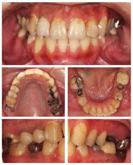
Figure 2: Pretreatment intraoral photographs.
Before starting comprehensive therapy, a thorough examination was conducted. Full mouth photographs and radiographs were taken. Dietary analysis and salivary risk assessments were obtained. Pretreatment records, such as diagnostic casts and diagnostic wax ups, were obtained and mounted on a semi-adjustable articulator. Examinations revealed high caries risk, according to CAMBRA [5]. Salivary assessment revealed a normal flow rate, medium buffering capacity, with high Streptococcus mutans and Lactobacillus bacterial counts [6]. The patient had poor dietary habits.
Extra oral examination revealed a symmetrical face with competent lips. The temporomandibular joint demonstrated a full range of function, without clicking or crepitus. Intraoral, a thin biotype gingival was observed [7]. Her Silness and Löe plaque and gingival index score was moderate [8]. Grade I gingival recession was found at tooth #12 [9]. During the dental examination, wearing facets were found, with a score of 0 (no wearing into the dentin) [10]. Teeth #18, 28, 38, 37, 47, and 48 were missing. Teeth #17, 26, 27, 36, 35, and 45 were non-restorable. Caries was found in tooth #16. Defective restorations were observed in teeth #15, 14, 12, 11, 21, 23, 24, 25, and 26. Periapical infections were present in teeth #15 and 14 (Figure 3). An endodontic diagnosis was performed following the American Association of Endodontists’ guidelines [11]. To plan and achieve a pleasing composition by multispecialty treatment, the patient’s smile was analyzed (Figure 4) [12]. The treatment alternatives, coupled with material selections, were discussed with the patient and informed consent was obtained.
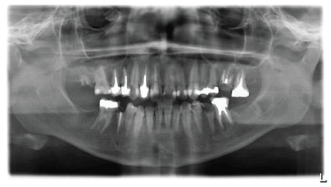
Figure 3: Pretreatment panoramic radiograph. Non-restorable teeth, multiple post and core restorations, and several preapical lesions can be observed.
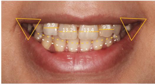
Figure 4: Preoperative smile analysis. Note the convex incisal plane following the lower lip, low smile line, absence of buccal corridor, slight deviation in the midline, and disproportionate anterior teeth.
Extensive prosthodontic therapy and a multidisciplinary approach were coordinated in order to obtain functional and aesthetic rehabilitation and smile improvement. As no emergency treatment was needed, treatment started with periodontal therapy that included scaling, prophylaxis, and fluoride application (Clinpro White Varnish, 3M ESPE, St Paul, MN). Teeth that could not be restored were extracted. Caries control was performed to eliminate active carious lesions and glass ionomer restoration was used for temporization (Ketac™ Fil Plus Aplicap™, 3M ESPE) [13]. Composite restoration was performed for tooth #16 using an A2 shade (Filtek Z350 XT, 3MESPE). Finally, the patient was encouraged to maintain good oral hygiene and the need for home care was reinforced.
Root canal treatment (RCT) was performed using a crown down and hydride technique for instrumentation [14]. Chloroform was used for dissolving gutta-percha points [15]. Furthermore, obturations were finished using a lateral compaction technique with AH plus resin sealer (Dentsply Maillefer, York, PN) and gutta-percha points (Figure 5) [16].

Figure 5: Preapical radiographs after RCT of teeth #13, 11, 21, 22, 25, and 34. An endodontic file was separated in the preapical area while treating tooth #25.
Teeth #21 and 25 had a composite core (Filtek Z350 XT, 3MESPE). Post space was prepared to receive fiber posts and cementation was performed with resin cement in teeth #13, 11, 22, and 32 (Relay X fiber post and Relay X unicem, 3M ESPE). Cone-beam computed tomography (CBCT) was performed and used to evaluate bone volume and quality. Apicoectomy was performed in teeth #15 and 14 (Figure 6). Additionally, to assess the implant position according to anatomical landmarks, a surgical stent was constructed [17]. Implant restorations were placed to replace teeth #26, 36, 35, and 45, using restorations of 5.0 × 8 mm, 4.3 × 11.5 mm, 3.5 × 11.5 mm, and 4.3 × 11.5 mm, respectively (Replace Select Tapered, Nobel Biocare, Kloten, Switzerland).
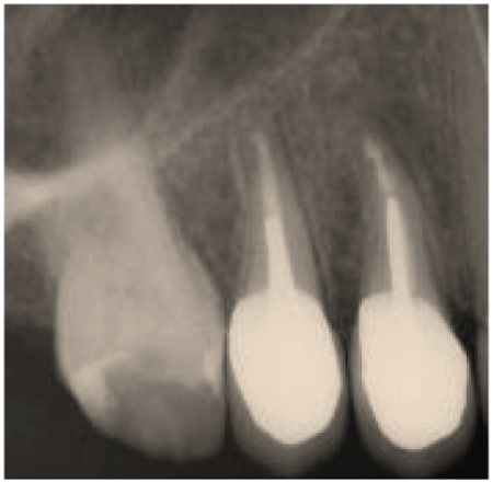
Figure 6: Periapical radiograph after apicoectomy. This procedure was performed to improve the prognosis of the preapical area as considerable periapical infection was present.
Full coverage restorations with a shoulder finish line were placed [18]. Moreover, laminated veneers with an incisolingual butt-joint design and a chamfer finish line, inter-proximally and cervically, were prepared [19]. Abutments were selected for implant restorations utilizing an open tray impression coping technique (Figure 7) [20]. Final impressions were taken using a custom tray with light- and heavy-bodied polyvinyl-siloxane impression material (3M ESPE). Temporary restorations were placed based on diagnostic wax up.

Figure 7: Radiographs showing the open tray impression coping technique at the abutment level.
Regarding material selections, IPS e.max Zir Pres (Ivoclar Vivadent, Schaan, Liechtenstein) was used to fabricate crowns at the posterior region; however, IPS e.max Ceram (Ivoclar Vivadent) was used for the other crowns and veneers. Porcelain fused to metal (PFM) was used for screw abutment implants and inserted according to the manufacturer’s instructions.
Restorations were tested to confirm that marginal discrepancy was absent and to verify the fit. Try-in pastes were used to check the color of the restorations. Crowns and veneers were cemented following the manufacturer’s instructions using resin cements (Relay X Unicem, 3M ESPE, and Variolink veneers, Ivoclar Vivadent, respectively). Implant screw holes were closed with tooth colored restoration (Filtek™ Supreme Ultra Flowable Dental Restorative Composite, 3M ESPE).
Follow-up and recall visits were planned every 3 months according to CAMBRA guidelines (Figure 8) [5]. The patient did not mention any complications and she was satisfied with her treatment (Figure 9).
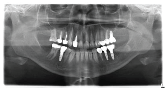
Figure 8: Post treatment panoramic radiograph.

Figure 9: Post treatment photographs. Note the bleach shade of the restorations.
A properly designed treatment plan is mandatory for overcoming challenges during rehabilitation treatment. Developing a systematic sequence and coordination among various specialists are required for complex prosthodontic work. The value of dividing treatments into a sequence of phases has been reported previously [3,4,21,22].
The goals for establishing objective rehabilitation are: freedom from disease throughout the masticatory system, maintenance of a healthy periodontium and teeth, a stable temporomandibular joint system, stable occlusion, functional comfort, and optimum aesthetics [23]. Gurrea, et al. reported that major disadvantages of the multidisciplinary approach are long-term appointments and patient acceptance. If a patient does not comply with one of the treatment phases, the ability to create the ideal result could be compromised [4]. However, in this report, all treatment options were presented to the patient. The first three phases were focused on controlling active caries and achieving disease stabilization. Special care was taken while dealing with her fragile and delicate gingival biotype [7]. Instrument breakage did not affect treatment prognosis and the patient was informed about this incident (Figure 10).

Figure 10: Preapical radiograph shows signs of healing after 5 months of follow-up. The presence of a separated file in the preapical area did not compromise healing.
The use of CBCT has been recommended for treatment planning of endodontic surgery (Figure 11) [24-26]. In 2014, Ee, et al. reported that a diagnostic accuracy rate of 76-83% can be achieved using such technology [27]. According to Misch, et al. single tooth implants have a 98.9% survival rate [28]. Prosthetic retrievability was the major reason for selecting screw-retainer restorations [29]. Moreover, an unfavorable ratio between the implant length and the superstructure, for instance, a short implant with a long crown, will not lead to pronounced crestal bone resorption [30,31]. In 2003, Goodacer, et al. reported that a major complication for ceramic restorations was crown fracture, which occurred especially in the posterior area [32]. However, with advances in dental ceramic material and adhesive systems, these materials now offer improved strength, retention, and better aesthetic control.
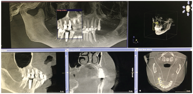
Figure 11: Preoperative assessment of the extent of the lesion using CBCT.
Dividing treatments into phases is essential to simplify complex prosthodontic work. A multidisciplinary approach is needed for managing such cases. Controlling the patient’s pain and anxiety, listening to the patient’s concerns, and explaining the facts about the treatment process can avoid discrepancy in outcomes.
This case was seen at Riyadh Elm University in Riyadh, Saudi Arabia as a requirement for the Saudi Board of Restorative Dentistry. This work would not have been possible without the guidance of instructors and colleagues at that institution.
Download Provisional PDF Here
Article Type: CASE REPORT
Citation: Alhekeir DF, Alsarhan RA, Alhekeir AF (2018) Treatment Phasing: A Simplified Approach for Complex Case Management. Int J Dent Oral Health 4(5): dx.doi.org/10.16966/2378-7090.278
Copyright: © 2018 Alhekeir DF, et al. This is an open-access article distributed under the terms of the Creative Commons Attribution License, which permits unrestricted use, distribution, and reproduction in any medium, provided the original author and source are credited.
Publication history:
All Sci Forschen Journals are Open Access