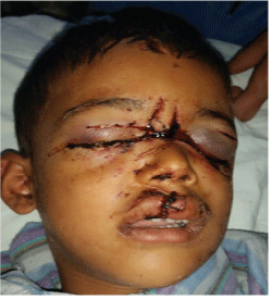
Figure 1: Pre- operative picture of the patient with dog-bite

Bhardwaj Y Arya S* Ram R Shinde A Ghezta N Sharma SK Ahsan R
Department of Oral and Maxillofacial Surgery, H.P Govt. Dental College & Hospital, Shimla, India*Corresponding author: Saurabh Arya, Department of Oral and Maxillofacial Surgery, H.P Govt. Dental College & Hospital, Shimla, Tel: +91-8894952773; E-mail: sourav.arya@gmail.com
Facial bite wounds in the maxillofacial region pose a unique challenge to the oral and maxillofacial surgeon. The main aim of the management is to neutralize any chances for infection and provide an infection free environment for wound healing. Most of the patients presented to us within 24 hours of the injury and we present our experience in restoration of esthetics and overall management of the patient. Use of prophylactic antibiotics, wound cleansing followed by primary closure was our treatment of choice.
Dog-bite; Wounds; Maxillofacial; Esthetics; Primary closure
Traumatic injuries to the skin due to animal bites constitute approximately 2 million cases per year. Dogs are responsible for approximately 60-80 % of such cases [1,2]. Superior extremities are afflicted in 45% of cases, lower limbs 25% and the head and neck region in 22% of situations, the face itself might be affected in 10% to 15% cases [3,4]. The surgical management of lacerations as a result of dog-bite remains an area of controversy among those who treat them. The factors affecting treatment options include - the nature of the injury, the expertise of the surgeon, the amount of time between injury and repair, and the location of the injury [5,6]. The primary closure of lacerations resulting from dog-bite injuries is still questionable. Some school of thoughts recommend allowing all animal bite injuries to heal by secondary intention whereas some suggest the primary closure of the lacerations.11 Oral and maxillofacial surgeons are commonly involved in the management of facial lacerations associated with dog-bite injuries. We present the management and treatment outcomes of 3 cases encountered at our center.
Subjects were enrolled from the out-patient clinics of oral and maxillofacial surgery. A retrospective medical record review was conducted of patients treated by the oral and maxillofacial surgery department between 2013-2016 for maxillofacial injuries. The data obtained from the patients’ medical records included patient demographics, event details, injuries sustained, and treatment rendered and analyzed. This is a retrospective study in which patients were assessed for the;
A 6 year-old male patient presented to the casualty with lacerations of the facial region (Figure 1). Affected individual had no neurological/ behavioral alterations associated with the injury as well as history of systemic illness and medication allergies. Primary assessment demonstrated that the patient was well oriented, hemodynamically stable and vital signs were within normal limits. Facial assessment exhibited a number of abrasions, a laceration relating to the upper labial region which went via the upper labial skin toward the mucosa. Two other lacerations involved the medial side of the eyes and extend to the infra-orbital rims as shown in figure 2. No bone or dental fractures were detected. Because of the patient’s young age, managing the injuries was difficult under local anesthesia. After proper facial disinfection and preparation of the operative field with sterile and clean surgical drapes, the wounds were meticulously irrigated with saline, wound debridement was carried out and sutures were attained under thorough care (5.0 polyglycolic acid absorbable sutures for the muscle together with oral mucosa; 6.0 nylon for skin) under general anesthesia. Long term follow up showed no facial nerve damage and patient was pleased with the esthetic outcome of the result.
Figure 1: Pre- operative picture of the patient with dog-bite
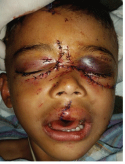
Figure 2: Post-operative picture of the patient with dog-bite
A 5 year-old female patient presented to the casualty with lacerations of the facial region (Figure 3). Primary assessment demonstrated that the Patient was well-oriented, hemodynamically stable and vital signs were within normal limits. Facial assessment exhibited lacerations in the bilateral cheek region. No bone or dental fractures were detected.
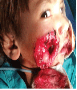
Figure 3: Dog-bite wounds in the cheek region bilaterally
A 52 year old patient reported with multiple abrasions and lacerations on the left side of the cheek (Figure 4,5 ). He was managed by primary closure under general anesthesia. Patient was pleased with the outcome of the surgery and there were no neurological deficits.
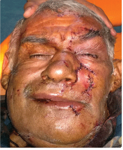
Figure 4: Elderly patient with dog- bite
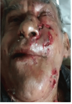
Figure 5: Management of the patient with primary closure
Primary evaluation was done for all the patients. Most of these patients were conscious, well oriented to time place and person, interacting well with our emergency team. Primary closure of the laceration was done under general anesthesia (Figure 6). Long term results show no facial nerve deficit and esthetically pleasing results (Figure 7).
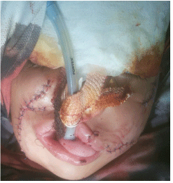
Figure 6: Oral intubation of the patient with dog-bite injuries
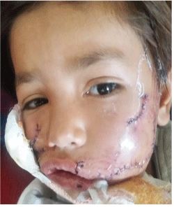
Figure 7: Primary closure under general anesthesia
After the administration of a local anesthetic all patients had their wounds copiously irrigated and cleaned with an antiseptic solution followed by saline. Primary closure on the day of presentation was achieved by layer by layer direct suturing. The repaired wound was left exposed to the air except in the cases where large wounds were present and rick of hematoma formation was there. Antibiotics were prescribed for seven days. Appropriate analgesics also were prescribed. Sutures were removed between 5 and 7 days postoperatively. Human rabies prevention was accomplished at the very same day and three days later Tetanus prophylaxis was also given in all the patients.
Results-A total of 3 patients were admitted with facial dog bites during the period studied which included mostly males. Several patients had multiple lacerations but each injured area has been recorded separately. Majority of the patients were less than 16 years old except for one patient who was 52 years old. Tissue loss was noted in 2 out of the three patients. Patients were followed up at 3 months, 6 months, 1 year and 2 years. We assessed the patient for facial nerve function and scar formation. Post- operative results showed no facial nerve damage and good esthetic outcome of the patients (Figures 8 and 9).
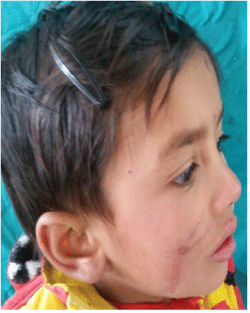
Figure 8: Follow-up of “case 2” (3 months follow-up)
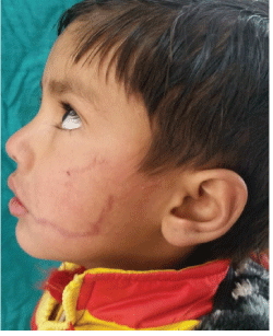
Figure 9: Follow-up of case 2 (3 months follow-up)
It is well known that a few hours after trauma the facial region is richly vascularised so proper wound management and primary healing are possible. When different tissues such as skin, muscle and oral mucosa are closed with appropriate suture materials, esthetically pleasing results can be obtained. The types of wounds encountered range from abrasions to severe facial injuries causing a severe psychological impact on the young. The children are most commonly affected by these animal inflicted injuries [7,8]. The tissue defects may be superficial or deep lacerations.
Most of these cases that presented to us received no treatment at the primary care centre even then primary closure was successful. Antibiotic therapy is indicated for infected bite wounds and fresh wounds considered at risk for infection, such as large wounds, large hematoma, full-thickness skin punctures and wounds with tissue loss of prophylactic antibiotics but there is no doubt that all dog bite victims should receive protective injections against tetanus and the possibility of contact with rabies should also be borne in mind [9,10]. Few authors have suggested that the surgical treatment be delayed and secondary intention wound healing in the management of such patients whereas many recommend primary closure [11-13].
Foreign body contamination may be associated with these wounds. Hence meticulous debridement must be followed by placing the patient on antibiotics. Penicillin derivatives form the mainstay of treatment in patients with dog bites [12,14]. We encountered foreign bodies in a number of patients and did debridement using saline, betadine and hydrogen peroxide.
Oral and maxillofacial surgeons are first ones to be called in the causality for the management of such crippling and debilitating injuries causing a severe psychological impact on the young. The aim must be to reduce scarring, prevent any nerve damage and meticulous debridement and closure under aseptic conditions and strictly following the anti-rabies protocol. Despite advice to perform delayed primary suture in extensive or difficult cases, all our patients had primary wound closure within a few hours of injury. We would strongly advocate primary wound closure for facial dog bites.
Download Provisional PDF Here
Article Type: Research Article
Citation: Bhardwaj Y, Arya S, Ram R, Shinde A, Ghezta N, et al. (2016) Current Perspective in the Management of Dog-Bite Patients - Our Experience in 3 Patients. Int J Dent Oral Health 2(5): doi http:// dx.doi.org/10.16966/2378-7090.199
Copyright: © 2016 Arya S, et al. This is an openaccess article distributed under the terms of the Creative Commons Attribution License, which permits unrestricted use, distribution, and reproduction in any medium, provided the original author and source are credited.
Publication history:
All Sci Forschen Journals are Open Access