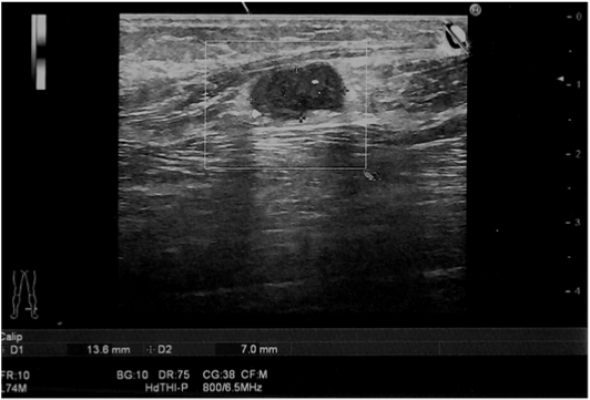
Figure 1: Ultrasonography scan of the external compartment of the left leg Presence of a well-defined, ovoid-shaped, homogeneously hypoechoic, lesion in the peroneus longus muscle.

Michaela Cellina1* Marcello Orsi1 Vincenza Fetoni2 Giancarlo Oliva1
1Radiological Department, Azienda Ospedaliera Fatebenefratelli e Oftalmico, Corso di Porta Nuova 23, 20121, Milan, Italy*Corresponding author: Michaela Cellina, Radiological Department, Azienda Ospedaliera Fatebenefratelli e Oftalmico, Corso di Porta Nuova 23, 20121, Milan, Italy, Tel: 0039- 0263632424; E-mail: michaela.cellina@fbf.milano.it
Schwannomas are encapsulated, benign, slowly growing tumors arising from Schwann cells of peripheral nerve sheath. Although they represent the commonest benign peripheral nerve sheath tumors, the occurrence on the lower limbs and especially from the superficial peroneal nerve is exceptionally rare. We present the clinical manifestations and imaging findings of a superficial nerve Schwannoma presenting as a painful mass in the external compartment of the calf, which was confirmed histologically and we correlate our experience with previously reported cases.
Schwannomas, also known as neurilemmoma, neurinoma, neuroma, nerolemoma or Schwann cells tumor, are well capsulated, normally benign, but occasionally malignant tumors, which originate from the Schwann cells in nerve sheaths with a slow-growing pattern [1,2]. They can be seen at any age, with a peak in patients 20 to 30 years of age1 and may occur singly or in multiple [1,2]. Most lesions are solitary and present as painless mass; neuropathic pain, weakness or dysesthesia can occur when the tumor grows large enough to compress the adjacent nerve [3]. Although they represent the most frequent benign peripheral nerve sheath tumors, and can affect any nerve within the body, their occurrence on the lower limbs is rare, accounting for 1% of all cases [4].
An otherwise well 50-year-old female, without any history of trauma, presented with a small mass in her leg on the outer side. She had noticed this mass 1 month before, without significant increase in size. She complained numbness in this region and local neurogenic pain. The pain was burning and intermittent, distally irradiated, worsened by local compression. Clinical examination confirmed the presence of a small (1 × 2 cm), well-defined, moderately tender lump in the lateral compartment of the left leg, deep in the peroneus longus muscle, with little mobility on the coronal plane. The pain did not increase with active or passive foot dorsiflexion. Neurological examination revealed a positive Tinel’s test: a slight percussion on the lump provoked mild tingling on the dorsal region of the foot. Motor function of the peroneal nerves was normal. No other lumps were palpable; no other pathologic finding was found.
To investigate this small mass, the patient underwent a left leg Ultrasonography (US) examination (Figure 1), showing an ovoid, welldefined, homogeneously hypoechoic nodule, then a Magnetic Resonance Imaging (MRI) with gadolinium injection (Figure 2), demonstrating a well-demarcated oval shaped lesion with homogeneous hyperintensity on STIR and T2 weighted images and homogeneous marked contrast enhancement, resulting in a diagnosis of suspected Schwannoma of the superficial peritoneal nerve. This diagnosis was confirmed by a tru-cut biopsy and immunostaining for the S-100 protein [5]. Surgical treatment was proposed and the patient is now waiting for enucleation.
Cases of Schwannomas arising from the superficial peroneal nerve are exceptionally rare: we found only 3 cases [6-8] reported in the Literature, one with no diagnostic imaging6, one with only MRI7, and one with only US [8].
The first report describes6 a 55-year-old male patient with a 6-month history of burning pain at his right fourth toe and at dorsal fourth web space, worsened pressing the mid-calf region. Physical examination of the foot was negative, but a palpable fullness in the proximal right leg was found. The patient underwent no diagnostic imaging, but a surgical exploration demonstrating a mass ensheating the superficial peroneal nerve. On histological investigation of the surgical specimen, a benign Schwannoma of the superficial peroneal nerve was found. Post-operatively his symptoms had completely abated. In this case, clinical manifestations are very different from those of our patient and quite atypical and no preoperative imaging examination was performed to study the mass.
Figure 1: Ultrasonography scan of the external compartment of the left leg Presence of a well-defined, ovoid-shaped, homogeneously hypoechoic, lesion in the peroneus longus muscle.
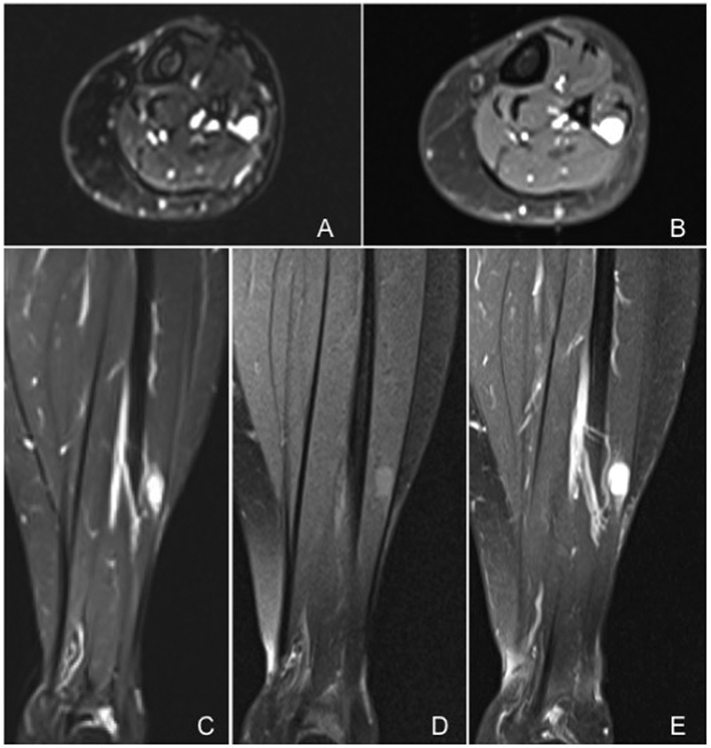
Figure 2: Contrast-enhanced MRI of the left leg Axial images: T2 weighted image with fat suppression (A) and T1 weighted image after contrast medium injection (B). Coronal images: STIR (C), T1 weighted image before (D) and after (E) contrast medium injection. Ovoid lesion with well-defined smooth margins in the peroneus longus muscle, hyperintense in basal acquisitions, especially in STIR and T2 weighted images, with a marked, homogeneous enhancement after contrast injection. After contrast medium injection (E) the continuity with superficial peroneal nerve, known as “tail sign”, is well visible.
The second one reports7 a 52-old female, affected by a gradually growing mass on her left leg, causing pain and numbness of the lateral aspect of her left leg for about 1 year; as in our case, the pain was burning and intermittent. The mass (3 × 2.8 cm) was investigated only with MRI, showing a tumor on the lateral leg compartment, ovoid in shape, with well-defined margins and strong homogeneous contrast enhancement, very similar to the images of our case. She was treated by surgical excision of the lesion and the histological analysis diagnosed a benign neurilemmoma with characteristics of ancient degenerative Schwannoma.
The last case [8] reports a 3-year history of two coexisting Schwannomas of the right ulnar and of the right superficial peroneal nerve, related to schwannomatosis disease, a rare form of neurofibromatosis, in a 63-yearold woman, who complained pain in her right limbs and numbness, weakness and paresthesias in the right forearm and hand. As in our case the superficial peroneal nerve Schwannoma presented as a small fusiform palpable mass (2 × 1 cm) and had the same US features of a well-defined homogeneously hypoechoic nodule. No MRI was performed before surgery.
In relation to its small size, the Schwannoma of our patient could be asymptomatic, however it was large enough to compress the adjacent nerve, causing pain. In the present case, imaging features of both US and MRI were typical for Schwannoma [9,10]; large tumors may exhibit cystic degeneration, calcification, hemorrhage and fibrosis (ancient Schwannoma) [10].
In summary, the interest of this case lies in the unusual location of the schwannoma: as far as we know, these neural sheath lesions rarely affect the nerves of the lower limb. In the differential diagnosis of soft tissue masses and nontraumatic leg pain, benign tumors, particularly Schwannomas of the peroneal nerves should be considered. They can be very difficult to diagnose clinically, and can often be mistaken for a lipoma: imaging studies, US and MRI, are helpful in achieving a correct diagnose.
Surgical resection with preservation of the nerve function is considered the treatment of choice [11].
This is a case report without any industry-sponsorship. Dr. Cellina, Dr. Orsi, Dr. Fetoni, Dr. Oliva report none to disclosures.
Download Provisional PDF Here
Aritcle Type: Case Report
Citation: Cellina M, Orsi M, Fetoni V, Oliva G (2015) Clinical Presentation along with Imaging Features of Superficial Peroneal Nerve Shwannoma: A Rare Case Report. J Neurol Neurobiol 1(3): doi http://dx.doi. org/10.16966/2379-7150.111
Copyright: © 2015 Cellina M, et al. This is an open-access article distributed under the terms of the Creative Commons Attribution License, which permits unrestricted use, distribution, and reproduction in any medium, provided the original author and source are credited.
Publication history:
All Sci Forschen Journals are Open Access