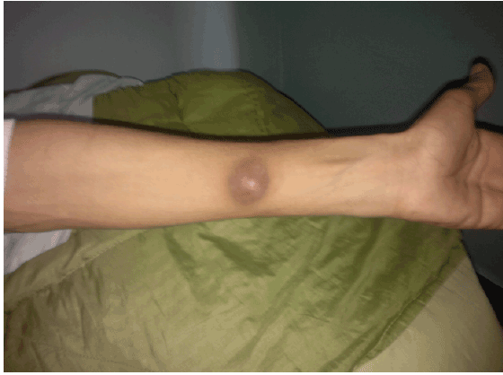
Figure 1:Tuberculous gumma

Benabdellah A1* Bachir N2 Belharane A2 Benabadji A2 Benchouk S2 Bensaha Z2 Brahimi H2 Lakhdori F2 Mahamdaoui F2 Taleb-Bendiab R2 Touati SN2 Labdouni MH3
1University of Oran, Ahmed Benbella, Algeria*Corresponding author: Benabdellah Anwar, HIV laboratory research, University of Oran, BP 1524 ELM Naouer 31000 Oran, Algeria, Tel: +213 (0) 41 58 19 47 /+213 (0) 41 58 19 41; E-mail: benabdellah.anouar@univ-oran.dz
The presence of cutaneous miliary tuberculosis in the AIDS era emphasized the importance of having a high index of suspicion for this condition in HIV-positive patients with skin lesions and advanced immunodeficiency. We report a 38 year-old male patient, diagnosed with HIV infection that developed disseminated tuberculosis in chest, abdomen and skin. While pulmonary symptoms improved under antituberculous drugs, skin lesion showed positive cultures for 6 months. He was healed after 12 months of treatment.
Miliary tuberculosis; AIDS; Tuberculous gumma
Tuberculosis has become more frequent since the emergence of HIV infection epidemics. Nowadays, there are important challenges, which complicate the management of HIV-TB co-infected patients. Among them, it is emphasized the increase of disseminated and extra-pulmonary tuberculosis forms, the multi-drug resistance and the increase in mortality.
A 38 year-old male patient was diagnosed 6 months ago with HIV infection. One month later he was admitted at the hospital with intermittent fever. At admission, the patient complained of asthenia, anorexia and important weight loss (5 kg), deshydratation and fever (39°). Crackles were audible over both pulmonary bases. The exam revealed hepatosplenomegalia and a red gummatous lesion of 4 cm of diameter developed on the internal surface of the left arm (Figure 1). Lymphadenopathy was absent and the blood pressure was normal. The laboratory tests showed a normal blood count (10 × 109 /l), hemoglobin 100 g/l, a high C reactive protein (100 mg/l; normal: 0-9 mg/l), liver tests and kidney function were normal.
Figure 1:Tuberculous gumma
HIV viral load was 40,000 copies/mm3 and a decreased CD4 cells (300/ mm3 ). Tuberculin test was negative. Acid-fast bacilli and culture results of sputum were negative.
A chest plain radiograph showed micronodular miliary. Thoracic MRI showed disseminated micronodular lesions. Abdominal MRI revealed ascitis, necrosed retroperitoneal adenopathies. The material collected from the skin lesion was studied for histopathology patterns, bacterioscopy and culture studies. Ultrasound confirmed a subcutaneous structural alteration on the internal surface of the left forearm with 04 cm in size. Skin histopathology studies revealed hyperacanthosis of the reticular dermis and mild infiltrate of lymphocytes around vessels and skin adnexia. Aspirated secretion from the lesion was negative for acid-fast bacilli. Culture studies revealed growth of mycobacterium tuberculosis.
Quadruple therapy with ethambutol, rifampin, isoniazid and pyrazinamide was started. After 3 months, there was a remarkable improvement in the patient’s general condition. Pulmonary symptoms improved under antituberculous drugs. Skin swabs showed positive cultures for 6 months. Two antibiograms did not disclose any resistance to tuberculostatic drugs. The patient was healed after twelve months of treatment.
Cutaneous tuberculosis is unusual in industrialized countries, most reports coming from developing countries [1,2]. The incidence of cutaneous tuberculosis has been reported to range from 0.15% to 0.26% [3]. Ravolamanana revealed that cutaneous tuberculosis occurred in 04.7% of extra-pulmonary manifestations of tuberculosis [4]. But the number of cutaneous tuberculosis observed in the north of Ethiopia, by Terranova [5], indicated a high incidence of the disease in the region. Terranova had identified 202 cases of cutaneous tuberculosis in the period of 34 months in the north of Ethiopia. 22% were HIV positive. Cutaneous tuberculosis is under diagnosed due to the low number of dermatologists and the poor life conditions in the population. Also, cutaneous tuberculosis diagnosis is difficult because of clinical polymorphism. Cutaneous manifestations are common in the HIV positive population and few lesions are biopsied [6]. The tuberculous gumma had been described during cutaneous tuberculosis. Kummar has found 05.4% of tuberculous gumma [2]. Terranova described 08.9% of tuberculous gumma. Assane found 11.25% of tuberculous gumma [7]. They had usually the aspect of cold abscesses. They are fluctuant, without painful. They were localized preferably on the members. Pulmonary (26.5%), osteoarticular (8.6%), neurological (0.66%) were the main visceral involvement [8]. Healing occurred after 1 to 2 months of treatment in all patients [9,10]. Recidivation occurred in 3 patients and 3 patients died [10].
Umapathy identified multidrug-resistance tuberculosis in 02% among 213 patients with cutaneous tuberculosis in India [9]. Cutaneous lesions usually respond favorably to antituberculous therapy; sometimes they require surgical management [10,11]. Slow responders to treatment have been reported, with lesions persisting occasionally >1 year. The recommended duration in cutaneous tuberculosis resulting from hematogenous spread is 10-12 months [12].
Cutaneous forms of miliary tuberculosis are a very rare subtype, resulting from a severe pulmonary infection overwhelming an already weakened immune system. Our patient presented lesions associated with concomitant clinical foci of tuberculosis make the diagnosis more evident. More common subtypes, scrofuloderma and lupus vulgaris, are more likely to be identified because of their frequency of occurrence.
Miliary tuberculosis has historically been extremely rare and well known for its occurrence in children; it is an increasingly serious infection in immunosuppressed patients, such as those infected with HIV, on long-term oral corticosteroid therapy, or on other immunosuppressive therapies for organ transplant or inflammatory or autoimmune conditions. Cutaneous skin lesions consist of small, erythematous to violaceous papules or pustules with hemorrhagic necrosis and umbilication affecting a substantial portion of the body. If healing occurs, lesions leave atrophic, depressed scars surrounded by a brownish, hyperpigmented halo. TST is typically negative because of anergy. Skin biopsy with histological examination reveals numerous microabscesses containing neutrophils and numerous mycobacterial organisms.
Metastatic tuberculosis abscesses (TB gumma) can arise from breakdown of an old healed tubercle that still contains live organisms or from cellmediated immune defense inhibition that reactivates. Tuberculosis gumma is usually seen in malnourished children and immunosuppressed adults. Single or multiple nontender, fluctuant nodules develop forming draining sinus abscesses unless surgically incised and drained. Nodules can occur at any location without any specific predominance. Histological examination reveals massive skin necrosis with copious mycobacterial organisms. TST is variable.
Our patient did not present the immune reconstitution inflammatory syndrome (IRIS). IRIS represents a diverse range of pathologic inflammatory manifestations that occur in the days to months after HAART is commenced. Many infectious and noninfectious dermatologic IRIS manifestations have been described. The common manifestations of Mycobacterium tuberculosis IRIS are new or worsening lymphadenopathy, pulmonary infiltrates, serous effusions, and neurologic involvement. Cutaneous involvement has been mentioned in three case series.
Cutaneous tuberculosis is quite unusual. HIV infection can favor dissemination and delay the microbiological and clinical responses. However, prolonged treatment with antituberculous drugs may be needed
Download Provisional PDF Here
Article Type: Case Report
Citation: Benabdellah A, Bachir N, Belharane A, Benabadji A, Benchouk S, et al. (2015) Cutaneous Tuberculosis in HIV Infected Patient. J HIV AIDS 1(2): http://dx.doi.org/10.16966/2380-5536.108
Copyright: © 2015 Benabdellah A, et al. This is an open-access article distributed under the terms of the Creative Commons Attribution License, which permits unrestricted use, distribution, and reproduction in any medium, provided the original author and source are credited.
Publication history:
All Sci Forschen Journals are Open Access