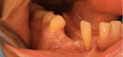
Figure 1: Preoperative photograph of the surgical site.

Raniah Abdullah Al Eid*
Department of Oral and Maxillofacial Surgery, College of Dentistry, King Saud University, Saudi Arabia*Corresponding author: Raniah Abdullah Al Eid, Department of Oral and Maxillofacial Surgery, College of Dentistry, King Saud University, Saudi Arabia, E-mail: dr.raniahaleid@hotmail.com
Impaction of mandibular permanent lateral incisor and canine is rare in dental practice, and its treatment is challenging because of their importance to facial esthetics and function.
Aim: This case report presents a rare deep impaction of permanent lateral incisor and canine with ectopic orientation of the teeth.
Material and Method: A 47 years old Saudi lady has been referred from her orthodontist to our Oral and Maxillofacial surgery clinic for consultation regarding the impacted mandibular right lateral incisor and canine. Investigations included cone beam computed tomography which showed that the right mandibular canine (#43) is impacted and the root is fully formed with dilacerations of the tip of the root, the crown is directed mesially. The right mandibular lateral incisor (#42) impacted lingual to impacted tooth #43 and the crown is directed distally below the apices of first and second premolar. Incidental finding of a unilocular radiolucency in the lower anterior area below the crown of impacted tooth #43. The treatment plan included surgical removal for future implant insertion while the orthodontic treatment was unapplicable in this case. Results: Patient undergoes surgical removal of the impacted teeth and enucleation of the associated cyst under general anesthesia.
Conclusion: Mandibular canine and lateral incisor impaction is a rare condition. Its treatment options are variable including no intervention, orthodontic and surgical removal. The selection of the treatment option is highly dependent on the patient interest and need and weighting them against the possible risks.
Impaction of mandibular permanent lateral incisor and canine is rare in dental practice, and its treatment is challenging because of their importance to facial esthetics and function. Tooth impaction can be defined as any tooth that fails to erupt into its normal position in the dental arch within the expected time and remains unerupted due to impediment at the path of eruption or abnormal tooth germ position [1]. The common etiologic factors are local (limited space for eruption, ectopic orientation of the tooth germ, supernumerary teeth, pathologic lesion, etc). Systemic and genetic factors are related to association with disorders or syndromes such as cleidocranial dysplasia and osteopetrosis [2]. Inheritance of dental impaction or delayed eruption as an isolated condition has been reported [3]. Dental impaction is a common problem occurring mostly in the following order of frequency: mandibular third molars, maxillary third molars, maxillary canine, mandibular premolar, maxillary premolar, mandibular canine, maxillary central incisors and maxillary lateral incisors. It is rare to see impactions of incisors and first molars [4]. Management of teeth impaction usually requires multidisciplinary approach including maxillofacial surgery, orthodontic, prosthetic rehabilitation and possibly periodontics.
Impaction of mandibular canines is uncommon and its incidence in the range of 0.10% to 1.29% [5-12]. Impaction of mandibular incisor has even much rare incidence which is in the range of 0.04% to 0.65% [13,14]. The impaction of both mandibular permanent canine and lateral incisor was reported only one time in the literature with our case as a second one, which indicates an extremely low incidence. There is recent evidence indicating a genetic interpretation of this condition [15].
There is an indication of the association between different dental anomalies. A cross-sectional study was performed to analyze the prevalence and associations between dental anomalies observable on panoramic radiographs in a large sample of non-orthodontic patients. Several associations was detected between different dental anomalies particularly, a significant association between supernumerary teeth and impacted teeth, tooth transposition; odontomas and impacted teeth [16].
This case report presents a rare deep impaction of permanent lateral incisor and canine with ectopic orientation of the teeth.
A 47 years old Saudi lady has been referred from her orthodontist to our Oral and Maxillofacial surgery clinic for consultation regarding the impacted mandibular right lateral incisor and canine for the possibility to do extraction and future implants. Patient is complaining from the poor aesthetic due to the space of lost teeth in the lower dental arch. No family history of tooth impactions.
Clinical evaluation revealed large space of missing teeth (impacted) in area of lower right canine and lateral incisor, decreased alveolar ridge height in area of the impacted teeth and drifting of the right central incisor distally with bone resorption and gingival recession (Figure 1).
Figure 1: Preoperative photograph of the surgical site.
The initial panoramic radiograph revealed impactions of the permanent mandibular right canine obliquely positioned with the crown pointed mesially and lateral incisor with a horizontal position under the first and second premolars and its crown is pointed distally.
For complete diagnosis, the patient was referred for cone beam computed tomography (CBCT) imaging with the following report was received: Cone beam computed tomography examination was performed using a medium field of view. Sectional images along multiple planes were obtained, including oblique cross-sectional images perpendicular and parallel to the jaws.
Reformatted cropped panoramic view shows that the right mandibular canine (#43) is impacted and the root is fully formed with dilacerations of the tip of the root, the crown is directed mesially (Figure 2).
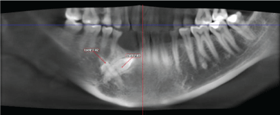
Figure 2: Reformatted cropped panoramic view of the impacted mandibular right canine and lateral incisor.
The right mandibular lateral incisor (#42) impacted lingual to impacted tooth # 43 and the crown is directed distally below the apices of first and second premolar , with this pathway of impaction it seems it will be difficult to exposes the tooth to be aligned in the proper location (Figures 4-6).
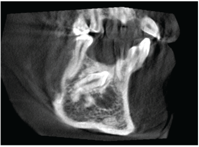
Figure 3: CBCT, sagital view showing impacted mandibular right canine and a unilocular cyst seen below its crown.
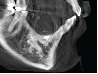
Figure 4: CBCT, sagittal view showing impacted right mandibular lateral incisor the crown directed distally.
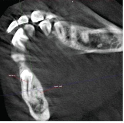
Figure 5: CBCT, axial view showing impacted right mandibular canine.
There is well defined unilocular radiolucency in the lower anterior area below the crown of impacted tooth #43. This could be OKC or simple bone cyst (Figure 3).
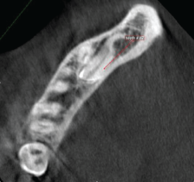
Figure 6: CBCT, axial view showing impacted right mandibular lateral incisor in almost horizontal position at the level of root apices of adjacent teeth.
After complete evaluation and orthodontic and periodontic consultation, the surgical treatment plan was set which included surgical removal of the impacted teeth which are the right mandibular lateral incisor (#41) and right mandibular canine (#43) with minimal bone removal as applicable. Associated cyst enucleation and histopathologic examination. Alveolar bone defect augmentation using guided bone regeneration technique. Two dental implants insertion after 4-6 months. Followed by prosthetic rehabilitation.
All treatment options, the treatment plan, the surgical steps, benefits, risks and possible complications were discussed with the patient. The patient preference was considered and she was consented before the procedure.
The surgical procedure was done in the usual standard manner under general anesthesia for patient convenience. Sulcular flap incision without vertical release was done. After buccal bone exposure the estimated position of the more superficial tooth which is the canine was pointed out. Canine exposed then removed smoothly. The lateral incisor was located very deep lingual and the two sockets were not connected leading to the need to remove additional bone. The impacted lateral incisor was then removed smoothly after sectioning the tooth. The small cyst located adjacent to them enucleated and the specimen fixed in 10% neutral buffered formalin for histopathologic examination. The resulted bone defect cleaned and repaired with guided bone regeneration using Small Geistlich Bio-Oss® granules (0.25-1 mm granules size) of 1 cc in amount which is a bovine derived xenograft. It was mixed in saline and adapted well to the defect walls then covered with Geistlich Bio-Gide® membrane size: 13 × 25 mm that consists of porcine collagen and has a bi-layer structure. The flap closed in water proof manner to protect the graft materials (Figure 7a-7f). Patient covered with broad spectrum antibiotic which is amoxicillin Clavulanate during the postoperative period for 5 days. During follow up appointments patient showed satisfaction and good healing progress with no signs of infection or wound opening (Figure 8a,8b). A long term follow up was scheduled for the patient and planned for delayed implant insertion after 4-6 months.
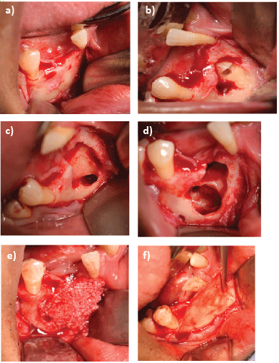
Figure 7: Surgical procedure.
7a: Flap reflection.
7b: Impacted canine exposure.
7c: Impacted canine removal.
7d: Impacted lateral incisor removal and the adjacent cyst lining.
7e: Bone graft for defect repair.
7f: Membrane adaptation over the bone graft.
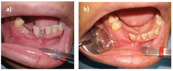
Figure 8: Postoperative follow up of surgical site.
8a: 2 weeks postoperative.
8b: 3 weeks postoperative.
The specimen of the cyst wall soft tissue was sent to histopathology laboratory. The microscopic examination reveals a section of fibrosed connective tissue with area of hemorrhage and mature bone fragment. The connective tissue has loose myxomatous to fibrous stroma and contains few mast cells and focal area of chronic inflammation in addition to islands of inactive odontogenic epithelial rest composed of clear cells that have swelling whorled morphology are seen. Interestingly, some of the epithelial rest cells contain brown pigment in their cytoplasm.
Histopathologic diagnosis: Connective tissue with inactive epithelial odontogenic rest and impacted lateral incisor.
As observed in literature there is no specific protocol for management of inverted impacted teeth varying from conservative management as a safest way to surgical removal with associated risks. The decision of surgical removal of the inverted impacted tooth might leads to complications such as damage to adjacent nerves and excessive bone loss due to great difficulty in surgical accessibility and aberrant position of the tooth [17,18].
Decision on the management protocol depends on the presence or absence of pathological changes in the tooth follicle and on the patient’s esthetics and functional needs. The management protocol of inverted impacted incisors must include prosthetic rehabilitation following surgical removal to restore aesthetic and speech.
Cozza P, et al. [19] reported a management of an impacted supplemental tooth similar in shape to lateral incisor in mandibular canine region in a mixed dentition aged boy. The management included extraction of the primary mandibular left canine early after detection to allow spontaneous eruption of the impacted tooth and then its extraction. A follow up period of 29 months post extraction of the impacted supernumerary tooth showed full eruption of the mandibular left canine into its normal position. This indicates that early management is mandatory to avoid complications at advanced age [19].
A case of a similar rare impaction has been reported [20]. The management included conservation of the impacted teeth using special tunnelling technique with ortho-surgical method. This method was inapplicable in our case due to the presence of a lesion associated with the impacted lateral incisor. According to Wetz if the canine crown migrated beyond the adjacent lateral incisor root apex, it is considered impossible to reposition the canine to its normal position orthodontically and if it is associated with pathology or neurological symptoms, surgical removal is mandatory [21,22].
In addition, the age consideration is an important factor in management decision. Our case is a late middle aged lady expected to be more difficult in manipulating such deep and complicated impactions with orthodontic techniques due to increased possibility of bone stiffness with older age. Moreover canine was dilacerated and the lateral incisor was located just apical to the adjacent premolars where orthodontic movement might be damaging.
In conclusion, mandibular canine and lateral incisor impaction is a rare condition. Its treatment options are variable including no intervention, orthodontic and surgical removal. The selection of the treatment option is highly dependent on the patient interest and need and weighting them against the possible risks.
The author has no conflict of interest.
Download Provisional PDF Here
Article Type: CASE REPORT
Citation: Al Eid RA (2018) Aberrant Impaction of Mandibular Canine and Lateral Incisor: a Case Report. Int J Dent Oral Health 4(4): dx.doi. org/10.16966/2378-7090.271
Copyright: © 2018 Al Eid RA. This is an open-access article distributed under the terms of the Creative Commons Attribution License, which permits unrestricted use, distribution, and reproduction in any medium, provided the original author and source are credited.
Publication history:
All Sci Forschen Journals are Open Access