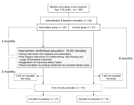
Figure 1: Flow chart showing the study protocol and participant selection procedure


Ayako Kubota1 Meiko Oki2 Yasuko Kawakami3 Kiyoko Kanamori3 Hiroji Shimomura4 Shiro Mataki1 Kumiko Sugimoto5*
1Department of Behavioral Dentistry, Graduate School of Medical and Dental Sciences, Tokyo Medical and Dental University, Tokyo, Japan*Corresponding author: Kumiko Sugimoto, Ph.D., Department of Oral Prosthetic Engineering, Graduate School of Medical and Dental Sciences, Tokyo Medical and Dental University, 1-5-45 Yushima, Bunkyo-ku, Tokyo 113-8549, Japan, Tel: +81-3-5803-4850; Fax: +81-3-5803-0237; E-mail: ksugimoto.bohs@tmd.ac.jp
Purpose: The increased prevalence of dental caries and gingivitis in the early 20s is a concerning oral health problem in Japan. The aim of the present study was to evaluate the effectiveness of a single individual education program regarding the oral health status and oral health behaviors for university students in their early 20s.
Methods: In total, 52 university students aged 20-22 years voluntarily participated in this study. Dental caries indices, the Plaque Index, the Community Periodontal Index-modified, and the unstimulated salivary flow rate were assessed at the beginning (baseline) and end (6 months after baseline) of the study. In addition, oral health behaviors were evaluated via a self-administered questionnaire at both time points. The intervention group received a single education session that included explanations of their oral health status, instructions on oral self-care, and suggestions on oral health behaviors 3 months after the baseline evaluation. The control group did not receive any intervention during the study.
Results: Twenty-seven students in the intervention group and 16 in the control group completed the study. The intervention group demonstrated an increase in the number of filled teeth and a decrease in Plaque Index scores at the end of the study. In addition, gingival bleeding did not exhibit further deterioration at the end of the study. These findings were not observed in the control group. The number of decayed teeth, pocket depth score, and unstimulated salivary flow rate remained unchanged at the end of the study in both groups. An increase in the flossing frequency and the practice of conscious brushing were observed at the end of the study in the intervention group, but not in the control group.
Conclusion: An individual education program for university students in their early 20s may contribute to improvement in their oral health status and behaviors.
Oral health status; Oral health behavior; Individual oral health education; Japanese university students
Retention of natural teeth is important for chewing and savoring food, and these functions are essential for maintaining holistic health and quality of life through the lifespan [1-3]. However, a high proportion of people lose their natural teeth because of various conditions, the most common ones being dental caries and periodontal disease [4]. The prevalence of untreated caries in permanent teeth is 36% in the total population, with a peak at the age of 25 years [5], while the prevalence of periodontal disease begins to increase at the approximate age of 20 years [6]. Deterioration of the oral health condition in young people in their early 20s is a concerning oral health problem in Japan [7], with a similar situation reported in several other countries [8-11]. Although acquiring good oral health behaviors and improving oral health status at this stage are crucial for the maintenance of oral health throughout life, deterioration of the oral health status and poor oral health behaviors may be caused by several factors in the young Japanese. Up to the high school level, the schools are obliged to conduct annual dental examinations by the School Health and Safety Act in Japan [12]. However, dental examinations are not mandatory for university or college students according to this regulation, therefore, most universities or colleges omit dental examinations from the annual physical examinations. Approximately 55% of individuals in Japan enter universities or colleges following graduation from high schools [13], and most of them do not receive regular dental checkups unless they have habits of regular dental checkups. In addition, the proportion of Japanese people in their 20s who received dental checkups in the past year was 29.1%, which was relatively lower than the proportions of Japanese people aged 20-60 years (35.7%) [14] and European people aged 15 years or older (50%) [15]. An irregular diet associated with the beginning of an independent life is another factor. Thus, young Japanese adults exhibit poor oral health behaviors and attitudes toward oral health, and intervention by dental professionals is particularly important for individuals in their early 20s. An individual instruction based on individual oral status was considered to be effective to improve individual oral health, because both individual and group oral hygiene instructions were reported to have similar beneficial effects [16].
Accordingly, to investigate appropriate interventions for the improvement of oral health in university students, an individual education session was conducted and its effectiveness was evaluated in the present study.
The participants were recruited from 160 students aged 20 years or older who were enrolled in a medical technology school in central Tokyo. These students were selected as participants because they were a uniform sample without any specialized knowledge or skills pertaining to oral health care. Before recruitment, the outline of the study was advertised to the students through posters placed on university notice boards. On the day of the annual physical examination, the students were informed details of the study, and 52 students voluntarily agreed to participate in this study and gave informed consents in writing. Students who were willing to afford the time to participate in a scheduled intervention session were assigned to the intervention group, while the remaining were assigned to the control group.
This study was conducted from April 2016 to October 2016. Oral examinations were conducted on the same day as the annual physical examination to evaluate the baseline status of the participants. Then, 3 months after the baseline evaluation, each student in the intervention group received a single education session that lasted for 15-20 min. The contents of the education program were determined for each individual by well-trained dental hygienists on the basis of the baseline assessment and the oral hygiene status on the day of the education session. The session basically included explanations of the caries and periodontal disease status, oral hygiene instructions for tooth brushing, daily flossing and usage of fluoridated toothpaste, and suggestions for improving dietary habits. The explanation of oral condition was conducted indicating the problematic parts with a hand mirror. Oral hygiene instructions included presentation of dental plaque, stained using a disclosing swab with a hand mirror, and demonstration and self-training of methods for tooth brushing and flossing.
In addition, participants with untreated dental caries were strongly recommended to undergo dental treatment. All participants were encouraged to continue self-care after the session according to the provided instructions. Six months after the baseline evaluation, oral examinations were performed again to evaluate changes in the oral health status. Questionnaire surveys regarding oral health behaviors were also administered along with the oral examinations at the baseline and end of the study (Figure 1).

Figure 1: Flow chart showing the study protocol and participant selection procedure
On the other hand, the control group did not receive any intervention during the study period. However, to avoid disadvantages due to the lack of an intervention, the participants were provided with explanations regarding their oral health status and instructions for oral health care after the evaluation at the end of the study.
This study was registered in the Clinical Trials Registry of the Center for Clinical Trials, Japan Medical Association (registration ID: JMA-IIA00282).
The oral examinations included the assessment of dental caries indices, the Plaque Index (PI), the Community Periodontal Index (CPI)-modified, and the unstimulated salivary flow rate. All examinations were conducted using an LED light in a classroom of the university.
Dental caries indices: The numbers of decayed, missing, and filled teeth due to dental caries, DMFT, were examined by trained dentists.
PI: According to the method proposed by Silness and Löe [17], four surfaces (buccal, lingual, mesial, and distal) of each target tooth (16, 12, 24, 36, 32, and 44) were examined and assigned a score from 0 to 3 according to the amount of plaque present. The score for an individual was obtained as the mean of scores for six target teeth which were calculated as the mean score for the four surfaces of each tooth.
CPI-modified: Measurement of CPI-modified scores was conducted basically according to the WHO Oral Health Surveys, fifth edition [18], except that the target teeth were confined to 10 teeth, same as in the fourth edition [19], considering population-based approach in the future. Briefly, the mouth was divided into sextants, and 17, 16, 11, 26, 27, 37, 36, 31, 46, and 47 were selected as target teeth. The examination was conducted in a bright room by well-trained dental hygienists using a WHO CPI probe, a disposable dental mirror, and an LED light. The depth of the gingival sulcus was measured with a force of less than 20 g. The highest pocket depth score for the target teeth was defined as the score for each sextant, and the highest score among sextants was taken as the individual score. Considering the age of the participants, attachment loss was not measured. Gingival bleeding was assessed separately, and the gingival bleeding status was evaluated on the basis of the number of sextants with gingival bleeding.
Unstimulated salivary flow rate: Unstimulated saliva retained in the oral cavity for 1 min was collected by the spitting method and immediately weighed.
A self-administered questionnaire was administered to all participants at baseline and the end of the study to evaluate changes in the oral health status and oral health behaviors after the intervention. The questionnaire was based on the FSPD-34 [20], with additional items added for the purpose of this study. The participants responded to 41 questions concerning oral health problems, oral health behaviors, and dietary habits (Appendix).
The intervention and control groups were compared at baseline using the Mann–Whitney U-test, chi-square test, or Fisher’s exact test. Changes in the oral health status and oral health behaviors at the end of the study relative to the baseline values in each group were analyzed using the Wilcoxon signed-rank test, the sign test, or McNemar’s test.
For further verification of the effects of the intervention, multiple logistic regression analysis was performed to assess changes in the gingival bleeding status at the end of the study. Prior to the analysis, independent variables were validated and a correlation coefficient of <0.8 was confirmed in order to avoid multicollinearity. Then, sex, salivary flow rate, PI score, maximum pocket depth score, frequency of interdental cleaning, frequency of sugar-containing beverage consumption, regular visits to a dental clinic, and daily brushing frequency at baseline were considered as adjusting factors. Smoking history was not included as an independent variable because of the small number of smokers.
The level of significance for all analyses was set at 0.05. All statistical analyses were performed using SPSS version 23.0J for Windows (IBM Japan, Tokyo, Japan).
This study was approved by the Ethics Committees of Tokyo Medical and Dental University (Approval number: 1170 in 2015) and Bunkyo Gakuin University (Approval number: 2015-0048).
In total, 29 and 23 participants were assigned to the intervention and control groups, respectively (Figure 1). Two and seven participants from the respective groups could not attend the evaluation at the end of the study and were excluded from the analyses. Eventually, 27 (15 men and 12 women; mean age, 20.6 ± 0.7 years) and 16 (6 men and 10 women; mean age, 20.8 ± 0.7 years) participants in the intervention and control groups, respectively, completed the study. Only one participant (3.7%) in the intervention group and two (12.5%) in the control group were smokers. The sex ratio and proportion of smokers were not significantly different between the two groups (P>0.05, chi-square test and Fisher’s exact test, respectively).
At baseline, 60.5% participants exhibited decayed teeth. The numbers of decayed and filled teeth as well as the DMFT showed no significant differences between the intervention and control groups at baseline (P>0.05; Table 1). While the number of decayed teeth was not changed at the end of the study in both groups, the number of filled teeth was significantly increased in the intervention group (P=0.037), with no change in the control group. In addition, the DMFT index was significantly increased in the intervention (P=0.007) and control (P=0.047) groups at the end of the study. There were no missing teeth at both time points in both groups.
| Intervention group (n=27) | Control group (n=16) | P-value Intervention vs Control at baseline | |||||
| Variables | Mean ± SD | Median (25th, 75th percentile) | P-value Baseline vs End of study | Mean ± SD | Median (25th, 75th percentile) | P-value Baseline vs End of study | |
| Decayed teeth | |||||||
| Baseline | 1.3 ± 1.4 | 1.0 (0.0, 2.0) | 0.9 ± 1.0 | 1.0 (0.0, 1.8) | 0.378 | ||
| End of study | 1.2 ± 1.2 | 1.0 (0.0, 2.0) | 0.630 | 1.3 ± 1.4 | 1.5 (0.0, 2.0) | 0.084 | |
| Filled teeth | |||||||
| Baseline | 5.1 ± 3.8 | 5.0 (1.0, 8.0) | 3.8 ± 3.8 | 3.5 (0.0, 6.0) | 0.244 | ||
| End of study | 5.9 ± 4.4 | 6.0 (2.0, 9.0) | 0.037* | 3.9 ± 3.8 | 3.0 (0.3, 7.5) | 0.642 | |
| DMFT | |||||||
| Baseline | 6.4 ± 4.4 | 7.0 (1.0, 10.0) | 4.6 ± 4.1 | 3.5 (1.0, 7.8) | 0.261 | ||
| End of study | 7.0 ± 4.7 | 7.0 (3.0, 10.0) | 0.007** | 5.2 ± 4.4 | 4.0 (1.3, 8.8) | 0.047* | |
| PI score | |||||||
| Baseline | 0.84 ± 0.50 | 0.79 (0.46, 1.21) | 0.59 ± 0.35 | 0.50 (0.31, 0.95) | 0.119 | ||
| End of study | 0.46 ± 0.28 | 0.42 (0.25, 0.67) | 0.001* | 0.71 ± 0.32 | 0.60 (0.47, 0.99) | 0.501 | |
| Unstimulated salivary flow rate (g/min) | |||||||
| Baseline | 0.554 ± 0.356 | 0.469 (0.237, 0.759) | 0.578 ± 0.269 | 0.477 (0.354, 0.869) | 0.514 | ||
| End of study | 0.582 ± 0.291 | 0.533 (0.332, 0.842) | 0.564 | 0.601 ± 0.326 | 0.530 (0.371, 0.751) | 0.717 | |
Table 1: Difference between the oral health status at baseline and that at the end of the study in the intervention and control groups
Differences between baseline and end of study values were analyzed using the Wilcoxon signed-rank test, while differences between the intervention and control groups at baseline were analyzed using the Mann-Whitney U-test.
**P<0.01, *P<0.05
The PI scores at baseline showed no significant difference between the two groups (P=0.119). At the end of the study, the PI score for the intervention group (P=0.001), but not the control group, exhibited a significant improvement (Table 1).
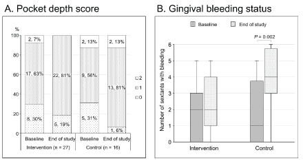
The pocket depth score at baseline was not significantly different between the two groups (P=0.896, Mann-Whitney U-test). Furthermore, there were no significant differences in the scores between baseline and the end of the study in both groups (P>0.05, sign test; Figure 2A). The number of gingival bleeding sextants at baseline was not different between the two groups, while that at the end of the study was significantly increased in the control group (P=0.002), but not in the intervention group (P>0.05; Figure 2B).

Figure 2: CPI-modified at baseline and the end of the study in the two groups
A. Proportion of participants with the highest pocket depth score of 0-2 (0: absence of condition, 1: pocket depth of 4-5 mm, 2: pocket depth of ≥ 6 mm) The number and percentage of participants are indicated in each bar. There is no significant difference between the two groups at baseline (P>0.05, Mann-Whitney U-test). Furthermore, there is no significant difference between the baseline and end of study values in each group (P>0.05, sign test).
B. Numbers of sextants with gingival bleeding at baseline and the end of the study There is no significant difference between the two groups at baseline (P>0.05, Mann-Whitney U-test). At the end of the study, the number has significantly increased in the control group (P˂0.01, Wilcoxon signed-rank test), but not in the intervention group (P>0.05).
As shown in table 1, the unstimulated salivary flow rate at baseline did not differ between the intervention and control groups and showed no significant changes at the end of the study in both groups.
The responses for the main questions pertaining to oral health and dietary habits are listed in table 2. At baseline, response rates for all questions except the one about interdental cleaning were not significantly different between the two groups. Response rates for the questions pertaining to the frequency of interdental cleaning and practice of conscious tooth brushing along the gum line were significantly different between baseline and the end of the study in the intervention group, but not in the control group. With regard to the other questions, there were no significant changes at the end of the study in both groups.
| Intervention group (n=27) | Control group (n=16) | P-value Intervention vs Control at baseline | ||||
| Percent of participants | P-value Baseline vs End of study | Percent of participants | P-value Baseline vs End of study | |||
| Q17: Do you regularly visit a dental clinic for checkups or tartar removal? | Options: yes, no | |||||
| Baseline | (18.5, 81.5) | (12.5, 87.5) | 0.695 | |||
| End of study | (25.9, 74.1) | 0.500 | (6.3, 93.8) | 1.000 | ||
| Q27: How many times a day do you brush your teeth? | Options: once, twice, three times or more | |||||
| Baseline | (3.7, 77.8, 18.5) | (12.5, 56.3, 31.3) | 0.731 | |||
| End of study | (0, 88.9, 11.1) | 1.000 | (6.3, 75.0, 18.8) | 1.000 | ||
| Q29: How often do you use an interdental brush or dental floss? | Options: never, occasionally, sometimes, frequently | |||||
| Baseline | (29.6, 55.6, 11.1, 3.7) | (62.5, 31.3, 0, 6.3) | 0.047* | |||
| End of study | (7.4, 48.1, 25.9, 18.5) | 0.013* | (62.5, 31.3, 6.3, 0) | 1.000 | ||
| Q30: Can you use dental floss or an interdental brush properly? | Options: no, not much, roughly, well, do not know | |||||
| Baseline | (14.8, 33.3, 25.9, 7.4, 18.5) | (31.3, 31.3, 6.3, 6.3, 25.0) | 0.468 | |||
| End of study | (7.4, 14.8, 63.0, 7.4, 7.4) | 0.359 | (37.5, 37.5, 6.3, 0, 18.8) | 1.000 | ||
| Q31: Do you try to brush between your teeth using the tips of the bristles of your toothbrush? | Options: no, yes | |||||
| Baseline | (33.3, 66.7) | (25.0, 75.0) | 0.735 | |||
| End of study | (11.1, 88.9) | 0.109 | (43.8, 56.3) | 0.375 | ||
| Q32: Do you consciously brush along the gum line when brushing your teeth? | Options: no, yes | |||||
| Baseline | (37.0, 63.0) | (25.0, 75.0) | 0.416 | |||
| End of study | (7.4, 92.6) | 0.008** | (37.5, 62.5) | 0.500 | ||
| Q33: Do you check your teeth and gums carefully with a mirror at least once a week? | Options: no, yes | |||||
| Baseline | (59.3, 40.7) | (68.8, 31.3) | 0.534 | |||
| End of study | (63.0, 37.0) | 1.000 | (81.3, 18.8) | 0.500 | ||
| Q41: Do you ordinarily take soft drinks or coffee and tea with sugar? | Options: often, sometimes, not much, rarely | |||||
| Baseline | (55.6, 18.5, 18.5, 7.4) | (18.8, 62.5, 12.5, 6.3) | 0.160 | |||
| End of study | (37.0, 25.9, 29.6, 7.4) | 0.424 | (25.0, 50.0, 12.5, 12.5) | 1.000 | ||
Table 2: Differences between oral health and dietary habits at baseline and those at the end of the study in the control and intervention groups.
Differences between baseline and end of study values in each group were analyzed using the sign test or McNemar’s test, while differences between the intervention and control groups at baseline were analyzed using the Mann-Whitney U-test, Fisher’s exact test, or the chi-square test.
**P<0.01, *P<0.05
Multiple logistic regression analysis for changes in the gingival bleeding status at the end of the study, which was adjusted for eight variables, revealed an odds ratio (OR) of 0.193 (95% confidence interval, 0.041-0.92) for the intervention group when the control group was used as the reference (Table 3). The gingival bleeding status was not significantly influenced by other baseline factors at the end of the study.
| Odds ratio | 95% CI | P-value | ||
| Group | Control | 1.00 (reference) |
||
| Intervention | 0.193 | 0.041-0.918 | 0.039* | |
| Sex | Female | 1.00 (reference) |
||
| Male | 0.687 | 0.134-3.516 | 0.652 | |
| Salivary flow rate | 0.692 | 0.060-8.003 | 0.768 | |
| Plaque Index | 0.329 | 0.040-2.671 | 0.298 | |
| Maximum pocket depth score | 2.353 | 0.601-9.216 | 0.219 | |
| Frequency of interdental cleaning | 1.576 | 0.527-4.708 | 0.416 | |
| Frequency of taking drinks containing sugar | 1.219 | 0.541-2.746 | 0.633 | |
| Regular visits to dental clinic | no | 1.00 (reference) |
||
| yes | 0.322 | 0.033-3.166 | 0.331 | |
| Daily brushing frequency | 0.812 | 0.181-3.651 | 0.786 |
Table 3: Multiple logistic regression analysis for changes in the gingival bleeding status in the control and intervention groups at the end of the study.
CI: Confidence Interval
*P<0.05
The aim of the present study was to evaluate the effectiveness of a single individual education session regarding the oral health status and oral health behaviors for university students in their early 20s. At baseline, 60.5% participants exhibited decayed teeth, and only 16.3% and 11.6% of them regularly visited dental clinics and practiced interdental cleaning, respectively. Moreover, approximately 30% participants exhibited new decayed teeth at 6 months from baseline. These results were similar to those reported by previous studies and surveys [7-9] in the sense that the prevalence rate of dental caries rises in the early 20s and provided further proof of the requirement for special intervention by dental professionals for individuals in this age group.
In order to investigate an appropriate intervention for university students, a single individual oral health education session was conducted in the present study. The evaluation at the end of the study demonstrated that this intervention was somewhat effective. In the intervention group, the number of filled teeth significantly increased, while that of decayed teeth showed no significant change. In the control group, the number of filled teeth was not significantly changed, while that of decayed teeth tended to increase. Moreover, the PI score was significantly decreased in the intervention group, but not in the control group. In addition, the results of the questionnaire survey demonstrated an increase in the frequency of interdental cleaning using dental floss and an improvement in the practice of conscious tooth brushing along the gum line only in the intervention group. Taken together, these results suggest that the single education session conducted by dental hygienists was effective in improving tooth cleaning skills and habits, resulting in a decrease in the PI score. However, the finding that the intervention group showed no change in decayed teeth with an increase in filled teeth implied that the present intervention was not sufficient for the prevention of dental caries. One reason for this result may be the unchanged dietary habit of consuming drinks containing sugar through the intervention, as a positive association between sugar-sweetened beverages or sugar intake and dental caries has been reported [21,22]. For the prevention of dental caries, continuous education on dietary habits to promote the acquisition of proper oral health behavior will be required.
Although the intervention group exhibited improved tooth cleaning habits at the end of the study, there was no significant change in the depth of the gingival sulcus. This suggests that the present intervention did not suffice for participants to acquire adequate tooth cleaning skills and habits, and that further professional care would be required to reduce the relatively deep periodontal pockets. The gingival bleeding status, another criterion for periodontal inflammation, also showed no significant change in the intervention group, while it significantly worsened in the control group. In addition, multiple logistic regression analysis adjusted for the oral hygiene status and oral health behaviors demonstrated that the OR for the gingival bleeding status decreased as a result of the intervention. This finding suggests that guidance regarding flossing and tooth brushing techniques prevented the worsening of gingival bleeding, although it was not sufficiently effective to improve the gingival condition.
Saliva plays an important role in maintaining oral health, with its various functions including moistening and lubrication of the mouth, protection of the oral mucosa, and protection of the teeth against dental caries [23]. In the present study, the unstimulated salivary flow rate was measured, and the baseline flow rate was not lower than that reported by a previous study [24]. Accordingly, any particular intervention to increase the salivary flow rate was not included in this program, which could have been a reason for the unchanged salivary flow rate at the end of the study, even in the intervention group. For an increase in salivary flow, further active programs, including massage of the salivary glands and tongue exercises may be required.
There had been few studies of intervention programs for oral health promotion among university students, though several studies had been conducted in younger school-going children [25,26]. Therefore, the present study was designed to assess the effectiveness of an intervention for university students who were busy with studies and did not have specialized knowledge and skills of oral health care. The finding that our intervention was somewhat effective may provide valuable information that can aid in the improvement of oral health in the general university student population. However, our study has some limitations. First, the participation rate for this study (about 33%) was relatively low, because the students were busy with clinical training and lectures and had difficulty in sparing time for the program. This observation suggests that the present participants may have had higher awareness about oral health than the students who were not interested in participating, and the oral health behavior of whole students might be worse than that implied by the present results. Thus, the small number of participants recruited from one university limited the ability to generalize the present findings to Japanese university students, although similar awareness level of oral health and oral health status have been reported for other Japanese universities [27,28]. In addition, it was not possible to randomly assign participants to the intervention and control groups because of the small number of participants. Accordingly, the intervention group comprised participants who were motivated to attend the intervention program or had some complaints regarding their oral health. The control group was presumed to comprise participants with fewer oral health problems. Second, only a single oral health education session was conducted for students in the intervention group because of their tight academic schedules. This could be one reason for the limited effects of the intervention. Third, it was difficult to incorporate the intervention immediately after the baseline evaluation because of the academic calendar; therefore, the intervention program was conducted 3 months after the baseline evaluation. Thus, it is possible that the oral health status may have worsened during the 3 months since the baseline evaluation in both groups, and that the final outcome for the intervention group could have improved more if the intervention had been implemented earlier.
To further understand the oral health status of university students, further investigations in a larger population and a variety of schools are required. In addition, more frequent interventions and further consideration of an intervention program suitable for university students are required to develop more effective oral health education programs.
Since the results suggested that individual intervention is effective for improving oral health of university students, providing oral examination and oral health education for every student by the support of university would be the ideal way to enhance the oral health of the students. However, such undertakings require considerable financial and professional resources and it would be difficult for most Japanese universities or colleges to implement them. Accordingly, a practicable approach may be to conduct questionnaire screening during the annual physical examination in universities and then encourage students to undergo dental treatments or checkups at their own expenses based on the questionnaire results.
A single individual oral health education session was implemented in university students with no specialized knowledge and skills regarding oral health care. The session included instructions pertaining to tooth brushing and daily flossing and suggestions to improve dietary habits for university students. At the end of the study, the number of filled teeth was significantly increased in the intervention group, but not in the control group. The PI score was significantly decreased in the intervention group and unchanged in the control group. The number of sextants with gingival bleeding was increased in the control group and remained unchanged in the intervention group. Furthermore, the questionnaire survey revealed an increase in the frequency of dental flossing and an acquisition of conscious brushing in the intervention group. Although participants were not randomized and the sample size was small, our results suggest that the present intervention was effective in improving the oral health status and oral health behaviors of the university students to a certain extent.
This work was partially supported by Collaborative Research Funding from the General Institute, Bunkyo Gakuin University. We deeply appreciate the contributions made by all students who participated in this study. The authors also thank Dr. Natsumi Tsuchihashi, Ms. Yoko Kono, and Ms. Rena Nakayama for their contributions to the implementation of the study and Ms. Naoko Adachi for her help with data analysis.
The authors explicitly state that there are no conflicts of interest in connection with this article.
Download Provisional PDF Here
Article Type: Research Article
Citation: Kubota A, Oki M, Kawakami Y, Kanamori K, Shimomura H, et al. (2017) Effectiveness of Individual Oral Health Education for Japanese University Students. Int J Dent Oral Health 3(5): doi http://dx.doi.org/10.16966/2378-7090.244
Copyright: © 2017 Kubota A, et al. This is an open-access article distributed under the terms of the Creative Commons Attribution License, which permits unrestricted use, distribution, and reproduction in any medium, provided the original author and source are credited.
Publication history:
All Sci Forschen Journals are Open Access