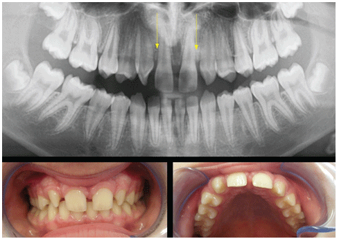
Figure 1: The case of the young male patient suffering from bilateral agenesis. The lack of space between central incisors and canine can be observed.

Marco Pasqualini1 Giorgio Comola2* Franco Comola3
1Private Practice, Milan, Italy*Corresponding author: Giorgio Comola, Universidad Alfonso X El Sabio, Madrid, Spain, Tel: 0039 3396897057; E-mail: drcomola@gmail.com
A miscellaneous method of both orthodontics and implantology is about to be shown by the authors. This technique is specifically suggested for the resolution of unilateral and bilateral agenesis of upper lateral incisors. One-piece immediate-load implants can be applied, after gaining the required space. Moreover, approaching this from an orthodontic angle, the main purpose is the restoration of the canine class (first class), which is crucial for both functionality and esthetics. The invisible aligners technique proves particularly useful in orthodontic distalizations, as it exploits each dental element, which is used as main anchorage. This procedure works by putting an increasing pressure only on the elements that are to be mobilized. The larger the teeth, the bigger the bearing surface which the aligner is able to exert these forces on. The invisible aligners technique combined with the responsible cooperation of the patient results in a comfortable and effective orthodontic treatment. The use of minimally invasive implantology originating from Italian School also allows the fast recovery of aesthetics due to immediate provisional crowns and it speeds the osseointegration process up with primary stability, as the bone quality is often poor in agenesis areas.
Immediate load: One-piece implants; Invisible aligners; Bicortical screw
The absence of one or more teeth in the anterior region might be easily noticed, causing aesthetic and functional disharmony. Agenesis cases of maxillary lateral incisors are quite common events [1,2]. On one hand, they are mainly an aesthetic issue to the patient, but on the other, a doctor’s first concern needs to be about occlusal-relationship alteration with possible scrolling by the protective canine guidance and the consequent chance to damage other dental elements [3].
In the case of a bilateral agenesis, an atypical increase of space between central incisor and canine can be observed at the time of permanent teeth development. In the case of unilateral agenesis, there is often a loss of the median line, which is always at the expense of the area where the tooth did not develop [4]. There are many options to treat congenitally missing anterior teeth.
For instance, the treatment which allows the closure of spaces by using auto-banding brackets and a preformed arch is frequently employed in orthodontics. By the procedure, it is possible to turn canines into lateral incisors by odontoplasty.
The first premolar is often turned into canine by using high-speed burrs, which minimize the palatal cusp, in order to make it a canine-like tooth. This way, the lateral disclusion is ensured [5,6].
As far as aesthetics is concerned, it might be regarded as a solution, yet this method does not take into account all the functional issues which may take place especially in unilateral agenesis.
The anatomy of a premolar does not actually fit the resistance to any of the strong forces exerted in lateral disclusions. Furthermore, the aesthetic performance requires a removal of dental material, which is invasive and irreversible solely for aesthetics [7].
Moreover, in this case, the propensity to third class suggests that spaces are being opened to improve the arch profile and to resolve the possible anterior crossbite; an aesthetic improvement in those patients with the concave profile is very frequent.
Despite the fact that the orthodontic opening of space is to be chosen for functional reasons, it might be contraindicated for some individuals, such as patients in first molar class with anterior overcrowding, in second class without any overcrowding, convex profiles with an excessive alveolar protrusion, since this kind of treatment would worsen their clinical condition and make their aesthetic features worse [8].
This restoration can be gained by using almost every unit fixed, although invisible aligners are more effective in the control of those very elements which need to be kept still. In addition, they prove to be more suitable for distalizations [9].
Furthermore, rehabilitation with minimally invasive one-piece implants, as indicated by Italian School, provides some other benefits. As far as any resistance to both static and dynamic strains is concerned, these implants are specifically suggested for immediate load and prove to be used whenever the bone appears slightly mineralized [10,11]. Bone thickness in the area of agenesis is typically inadequate, for the tooth fails to develop. Nonetheless, if the eruption process leads the canine to erupt nearby the lateral incisor, a better preservation of the good features can be noticed in the trabecular bone. There is evidence of quick bone reabsorption in those edentulous areas which originate after the development of permanent teeth when this latter one does not actually exist.
Some authors suggest keeping deciduous teeth in a temporary position in order to ensure a decent quality of the bone, once the patient is old enough to go under implant-prosthetic surgery [12,13].
Immediate-load one-piece implantology by Italian School is preferable because of its quick clinical effectiveness. It is, indeed, strongly suggested when there are signs of anatomical thinness or there is not much bone available; it is preferable also when spaces between canine and central incisor are small or high-impact reconstructive surgery, such as couplings, onlays, and membranes, is not to be chosen.
These implants do not need particularly large spaces since their diameter is between 2 and 3.5 mm in the spire and take advantage of both occlusal and deep cortical (Bicorticalism) [14,15].
Invisalign® method is aimed to gain more orthodontic space. Initially, the first series of these aligners was produced out of 4G polymer. Subsequently, since 2012, the new Smart Track material has been employed.
In the first case about unilateral agenesis, the “Invisalign full” procedure was used right before two refinements and the final restraint. On the other hand, in the second case about the lack of both incisors, a midcourse correction proved to be necessary.
Some attachments, made out of composite resin were applied, in order to improve the aligners. This method, in fact, allows exerting a continuous and increasing pressure, so that it is possible to keep the aligners steadier, which, moreover, simplifies every change of position.
As far as implant-prosthetic surgery is concerned, it was decided to use bi-cortical Garbaccio® screw type one-piece implants, made out of titanium. Their diameter in the spire is between 3.2 and 4.5 mm, while, in the core, it is between 2.2 and 2.5 mm, according to CE rules (0434). The length of the implants needs to be variable as well. They are, in fact, as long as it takes to get to the deep cortex.
The preparation of the implant site is performed with the flapless method, by using Pasqualini self-centering burrs, which have an increasing diameter (1.1-2.5 mm) and are assembled on a micro-engine, cooled by normal saline [16].
More specifically, the procedure was started with a probe-type burr (1.1 mm diameter), which is used to perform the early osteotomy and also makes it possible to sense the typical change of bone density in the deep cortex.
Right after radiographic tests, the very same length used earlier to set the probe-type burrs is employed again in self-centering burrs, in order to prepare the implant site. Besides, self-centering burrs are equipped with a triangular sharp head, whereas they appear to be blunt backward. This main feature enables accurate surgical tunnels to be performed, resulting in a minimally traumatic event to the receiving bone. Once the surgery is done, this technique makes it possible to use an immediate-load with a fixed prosthesis made out of resin, which is a polycarbonate ION crown, directly set on the stump.
According to this method, the patient is supposed to be anesthetized with a simple plexus anesthesia and treated preventively with antibiotics, such as amoxicillin and clavulanic acid 2 gr. per day for five days, and, if need be, with an anti-inflammatory like ketoprofen.
The immediate-load method is performed first by using polycarbonate and acrylic resin crowns, and secondly, after 60 days, once the osseointegration is completed, by applying definitive prosthesis made out of metal-ceramic [16].
A multicentric study about this implantology technique was carried out by six different private practices all over Italy (Busto Arsizio Varese, Milano, Como, Venezia, Bergamo, Roma). 62 patients suffering from both unilateral and bilateral agenesis have been under study between 2011 and 2016, with a 5-year follow-up.
The research was carried out according to the ethical parameters listed in the Declaration of Helsinki. In addition, each patient signed an informed consent, before being recruited.
Since no lack of osseointegratoin was observed, it was decided to consider as a failure every time the patient complained about aesthetics (Table 1).
| Unilateral | Bilateral | Rate | |
| Male (22) | 12 | 10 | 95.40 |
| Female (40) | 26 | 14 | 97.50 |
| Total (62) | 38 | 24 | 96.47 |
Table 1: Summary table of the 62 patients treated with invisible aligners and immediate-load one-piece implants.
This report is about a 13.y.o. male patient, Caucasian, suffering from agenesis of both lateral incisors in bilateral class one molar (2.1 1.2) in good health status. He showed a deep bite, resulting in a remarkable reduction of transverse diameters in the central group.
The above pictures prove, therefore, how the orthodontic Invisalign® treatment ensured to achieve more space. In this very case, the therapy lasted from May 2011 until April 2014, using overall 90 aligners gaining a total of 4,9 mm as space for the 1.2 and 4.8 mm for the 2.2.
The edentulous condition is characterized by little available space, in addition to remarkable bone atrophy. In order to restore the missing tooth, the procedure, previously described, was performed by employing, eventually, noble-metal alloy, and ceramic (Ivoclar Vivadent InLine®) crowns (Figures 1-8).
Figure 1: The case of the young male patient suffering from bilateral agenesis. The lack of space between central incisors and canine can be observed.
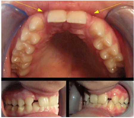
Figure 2: The space gained after the orthodontic treatment in order to insert conservatively the implants.
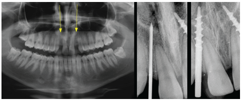
Figure 3: The radiographs: 1 that shows the remaining bone volume. The Pasqualini self-centering burr, which allowed to make an osteotomic tunnel specific for the implant insertion. Two bicortical screws (3.5 mm in the spire) at the end of the procedure.
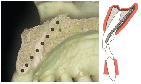
Figure 4: Anatomical section and schematic illustration of the way used to insert this kind of implant. By using this technique, the stump of the monophasic implant can be parallelized with the adjacent teeth directly in the mouth, right after considering the inclination- Titanium grade 2 can be bent with a forcep in order to achieve the right paralelism.
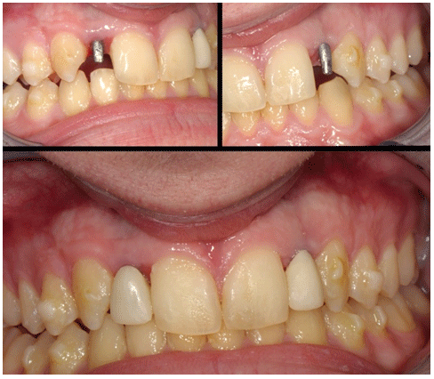
Figure 5: Two parallelized implants, applied with flapless technique. Downwards two immediate provisional elements at the end of surgery.
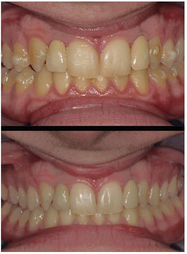
Figure 6: Two metal-ceramic crowns while being cemented. Downwards the same ones after two months.
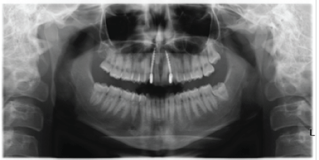
Figure 7: X-rays show two implants properly set in the bone, which appears to have minimal thickness.
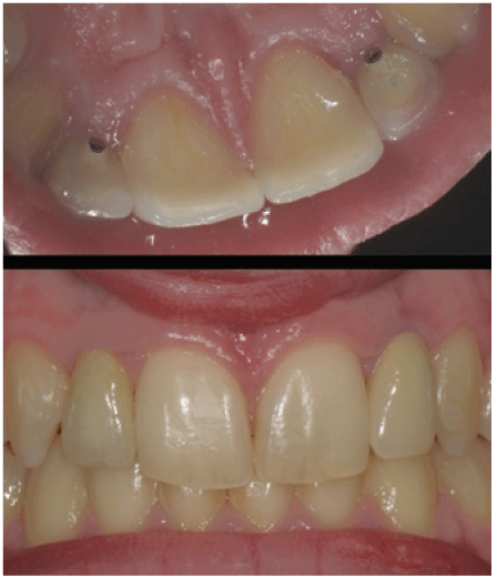
Figure 8: 18 months Follow-up. The features of both parietal and vestibular mucosa can be seen. Healthy gingival tissue and good esthetic contours are present at the present follow-up.
The second report deals with a 28-y.o. female patient, Caucasian, in a good health status, suffering from agenesis of the upper lateral left incisor, in bilateral class one, with a right canine mesialization, which affected the natural lateral space closing it.
The patient was treated with Invisalign® invisible aligners from September 2007 until July 2008, using overall 25 aligners. The pictures above show the results obtained by the orthodontic treatment with 4.6 mm of space gained for the lateral incisor. On July 15th, 2008, she underwent an implant procedure, after that an immediate acrylic provisional crown was placed during 2 months. After that, a definitive crown made of the same materials as stated before was placed and has been regularly followed-up between 2007 and 2016 (Figures 9-14).
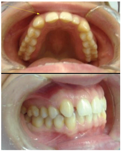
Figure 9: Lack of the upper lateral tooth in a young female patient.
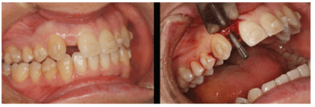
Figure 10: Gaining space allowed to set the bicortical screws with a small opening flap.
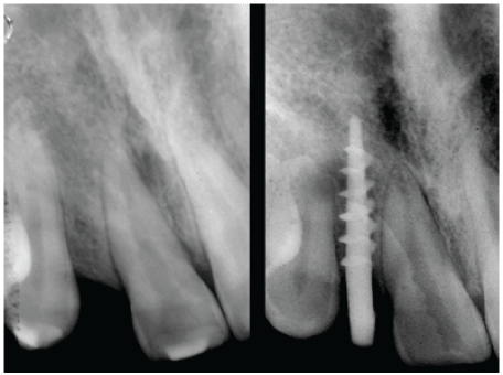
Figure 11: Peri-apical radiograph before and after the placemente of the dental implant.
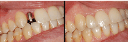
Figure 12: The modeling of the mucosa around the implant after removing the provisory cemented element at the end of the procedure. Downwards the definitive tooth made out of metal-ceramic.
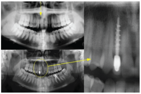
Figure 13: Radiographs of the case.
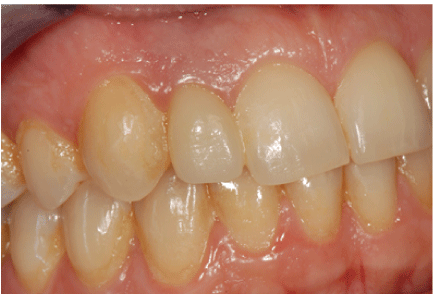
Figure 14: The crown of the conservative implant in the lateral agenesis. 8-years follow up.
Based upon obtaining information as requested in the case reports a discussion on traditional non-bicortical on Garbaccio style implants should be discussed as an alternative point of view. In light of the clinical outcomes, also proved by the pictures, it can be asserted that one-piece implants successfully allow reaching the given therapeutic goals [17,18]. In fact, although these implants look very thin, they prove to be extremely effective to treat agenesis in the maxillary areas, where the bone quality often appears particularly poor. Standard biphasic implants generally have a diameter of about 3.0 mm, nevertheless, considering the guidelines shown in other studies, the minimal bone mass around an implant is supposed to be at least 1.0 mm [19]. This measure means that the bone between a central incisor and canine has to be about 5.0 mm wide; an aim which is hard to reach, as the spaces obtained by performing orthodontic treatments are often very small. That is why both Italian and standard international implantology acknowledge that the usage of small implants on one hand preserves the alveolar bone from its physiological reabsorption and on the other ensures better aesthetic and functional results [20].
The immediate primary stability of Italian School monophasic implants makes the essential condition for the success of this procedure. This is made possible by both bicorticalism and adhesion to inner and/or outer cortex.
The implant insertion, in fact, often proves to be a challenge to implantologists, whenever bone spaces are small. Nonetheless, the procedure we suggest makes it possible to solve these problems by using thin monophasic implants, which ensure a lasting mucosal aesthetic, after being parallelized directly in the mouth [21].
These implants may be inserted obliquely in order to cover as much bone surface as possible. Moreover, thanks to the features of titanium (second grade) [22], they allow the operator to perform the parallelism when the adjacent teeth are still in the mouth with a manual folding simply using a forceps and, besides, at the very end of the procedure they ensure a direct insertion of a provisional, made out of acrylic resin or other provisional material. Of course, this final procedure is performed respecting the anatomy of the coronal portion of the dental implant. What is more, this orthodontic technique makes sure to effortlessly obtain a good distalization of the dental elements. It is essential, though, to pay attention to the torque, when trying to obtain radicular space, which is where the implants must be placed [23-25].
The importance of treating a collaborative patient deserves to be pointed out. In fact, the aligners require being used at least 22 hours per day in order to maintain their effectiveness.
Patients have the chance to foresee their aesthetic improvement, thanks to the insertion of some elements made out of composite resin (Pontic) within each empty space. Then, implants will be placed in these very areas, right after gaining a suitable space.
Furthermore, after the insertion of definitive crowns, it is crucial to keep working in order to preserve the results of the procedure. Therefore, patients are equipped with retainers (Vivera), which have to be used every night during the first six months. After this period the patient can reduce the frequency, using retainers from three times a week to just one night every ten days. Therefore, this progressive reduction looks like the only way to ensure lasting results [26].
The immediate load implantology technique with a small diameter is particularly suggested for a genetic monoedentulia, mostly when the aesthetic condition requires prompt action
It is necessary, though, to take into account some of the features of the frontal teeth (central and lateral incisors and canines), because they show a different inclination from that of premolars and molars [27].
On one hand, occlusal force vectors are not coaxial with the main axis of the roots, but rather they exert transversal forces. On the other hand, those belonging to molars and premolars cause forces that dissipate along the main axis. This is why frontal teeth must not have any contact in maximum intercustpation. Otherwise, they would be severely damaged in the last stage of deglutition (over-occlusion) by transversal forces which are not coaxial with the main axis of the roots. Because of this mechanism, even prosthetic crowns placed on anterior implants, as well as their matching teeth, need to be free from any static contact [28].
The importance of a proper occlusion reflects in the length of the rehabilitation, as an armonic occlusion can lead to a long lasting result. Unfortunately, a possible failure is often recognized due to microbiologic complications, systemic pathologies, inappropriate hygiene or smoking, while it is necessary to pay attention to the occlusion [29,30].
Download Provisional PDF Here
Article Type: Case Series
Citation: Pasqualini M, Comola G, Comola F (2017) Agenesis of Maxillary Lateral Incisor Treated with Orthodontics and Mini Dental Implants the Combined Technique with Invisible Aligners and Minimally Invasive Implantology of Italian School. Int J Dent Oral Health 3(4): doi http://dx.doi.org/10.16966/2378-7090.238
Copyright: © 2017 Pasqualini M, et al. This is an open-access article distributed under the terms of the Creative Commons Attribution License, which permits unrestricted use, distribution, and reproduction in any medium, provided the original author and source are credited.
Publication history:
All Sci Forschen Journals are Open Access