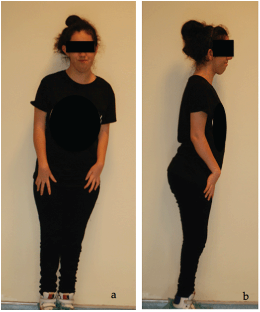
Figure 1: General appearance of an artrogryposis patient a: frontal view; b: lateral view.

Burcu BAL1* Nurhan GULER2
1Assistant Professor, Department of Prosthodontics, Faculty of Dentistry, Yeditepe University, Istanbul, Turkey*Corresponding author: Assist. Prof. Burcu BAL, Department of Prosthodontics, Faculty of Dentistry, Yeditepe University, No:238, Bagdat Cd, 34728 Goztepe, Istanbul, Turkey, Tel: +902163636044; E-mail: drburcubal@gmail.com
Mandibular hypomobility results from various disorders affecting the stomatognathic system. Arthrogryposis multiplex congenita (AMC) is an uncommon systemic disorder with mandibular hypomobility involved. Notably few cases of AMC with limited mouth opening and reduced range of jaw movement have been described in which this restriction has been attributed to involvement of the coronoid process. The cases diagnosed as AMC with mandibular hypomobility caused by coronoid hyperplasia are presented to highlight the clinical and three dimensional cone beam computed tomography findings of AMC patients.
Artrogryposis multiplex congenita; Coronoid hyperplasia; Mandibular hypomobility
Mandibular hypomobility is a challenging disorder that leads to restricted mouth opening, ranging from a partial reduction to complete immobility of the jaw [1]. The associated problems involve food intake, mastication, facial appearance, speech development, dental and oral hygiene, dental treatment and difficulty with intubation caused by airway malfunction [2,3]. The factors affecting mandibular hypomobility are caused by intraarticular disorders, such as the following: temporomandibular joint (TMJ) internal derangement (arthritis, dislocation); TMJ pathologies and extra-articular disorders secondary to dental injections, including trismus; fibrosis following radiotherapy; tumors involving the head and neck region; postsurgical ankylosis; postoperative problems (cranial base manipulation, orthognathic surgery); infections (otitis media, mastoiditis, ascending odontogenic formations, fascial space); traumatic injury and extracapsular fracture (condylar head, coronoid process, zygomatic arch); coronoid neoplasia/hyperplasia, myofascial pain dysfunction, rheumatologic disorders (osteoarthritis, rheumatoid arthritis, still disease, ankylosing spondylitis, scleroderma); and arthrogryposis multiplex congenita (AMC) [3-6]. Arthrogryposis multiplex congenita (AMC) is a rare disorder observed in approximately 0,03% of newborn infants with a systemic condition where mandibular hypomobility is involved [7]. The population prevalence of AMC ranges from 1/3300 to 1/56000 [8].
Arthrogryposis multiplex congenita is a nonprogressive syndrome characterized by congenital joint contractures occurring in two or more joints in different body areas [7]. AMC could be caused by myopathic or neuropathic disorders or other factors which decrease fetal movement [8]. Six major causes have been postulated for decreased fetal movement; fetal neuropathic abnormalities, fetal muscle abnormalities, fetal connective tissue abnormalities, space limitation (oligohydrodinamnios, multiple pregnancies), intra uterine vascular compromise and maternal disease [9,10]. A comprehensive evaluation such as history of pregnancy and delivery, family history, physical and neurological examinations, involvement of affected joints, intellectual development, and response to treatment is necessary for the diagnosis of arthrogryposis. Laboratoty tests, such as electrophysiological studies, pathologic examinations and gene sequencing may help differential diagnosis [11]. Prenatal screening which shows lack of mobility and abnormal position of the joints has become more widely available. 75% of AMC is not diagnosed prior to delivery and only severe AMC may be discovered by ultrasound screening [12]. Associated contracture and immobility of a tendon could cause further contractures and reduce mobility of the joints [13]. The upper and lower extremities are usually involved. Some of the more common signs and symptoms are associated with the shoulders (internal rotation), elbows (extension and pronation), wrists (volar and ulnar), hands (fingers in fixed flexion and thumb-in-palm), hips (flexed, abducted and eternally rotated, often dislocated), knees (flexion), and feet (clubfoot). Patients might have complications including scoliosis, pulmonary hypoplasia, respiratory problems, growth retardation, and abdominal hernias [14].
Hemangiomas of the upper face, low-set ears, a short neck and expressionless face are extraoral findings. Patients with AMC might have a micrognathia, cleft palate, high-arched palate, tongue with limited protrusion, dysphagia, weak masticatory and laryngeal muscles, poor gag and sucking reflex and microstomia [15-17]. Although nearly every joint is involved in severe cases of AMC, the TMJ is affected in 25% of AMC patients with limited mouth opening caused by mandibular hypomobility [17,18].
Notably few cases of AMC with limited mouth opening and reduced range of jaw movement have been described in which this restriction has been attributed to involvement of the coronoid process [15,16,19-22]. In this report, two cases diagnosed as AMC with mandibular hypomobility caused by coronoid hyperplasia were presented with clinical and three dimensional cone beam computed tomography (3D-CBCT) findings.
A 13-year-old girl diagnosed at birth with AMC was referred to our faculty with a toothache as the chief complaint. The patient had been too many dental hospitals, but treatment could not be performed because of limited mouth opening. Since then, she had occasionally used antibiotics without any dental treatment. The medical history revealed that she had undergone multiple surgical procedures associated with hip dysplasia and wrist treatments. No contributory family and social history was reported.
The extra oral examination showed obvious deformities of the limbs, particularly of the knees, causing difficulty to take a step. Postural alteration of the head, shoulders and feet were observed (Figures 1a and 1b).
Figure 1: General appearance of an artrogryposis patient a: frontal view; b: lateral view.
Asymmetry on the left side of the face and an abnormal skeletal relationship of the upper and lower jaws were observed (Figures 2a-2c).
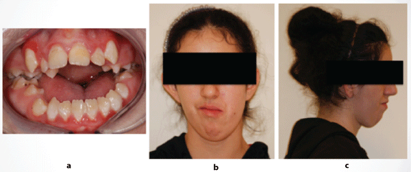
Figure 2: Clinical views of the patient a: frontal view of the mouth; b: frontal view; c: right lateral view.
The lateral and forward movement of the mandible was restricted. The translation of the condyles could not be detected, and there was no pain during the function and palpation of the masticatory muscles. During assisted opening, there was a hard end feel. The patient had a 10 mm open bite with only the second molars in contact. The maximal mouth opening measured between the incisal edges of the maxillary and mandibular left incisors was 20 mm. The intraoral examination showed poor oral hygiene and periodontal problems (Figures 3a and 3b).
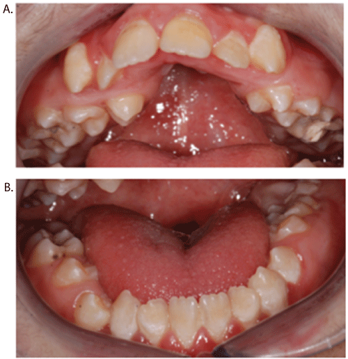
Figure 3: Severe marginal inflammation and extensive calculus and plaque may be observed. A: occlusal view of the maxillary arch showing micrognathia and high arched palate B: occlusal view of the mandibulary arch.
There were malpositioned teeth within and between each arch and had high arched palate. A carious lesion was detected in the left maxillary first molar. The panoramic radiograph showed a bilateral short ramus (Figure 4).
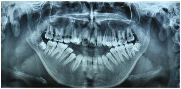
Figure 4: Panoramic radiograph at initial examination.
The antegonial notch was accentuated. With three-dimensional cone beam computed tomography (3D-CBCT) (NewTom 3G, QR, Ferrara, Italy), no anterior translation was observed upon mouth opening which confirmed the possibility of enlargement of the coronoid process (Figures 5-7).
The dental treatment involved an attempt of vital amputation for the left maxillary first molar diagnosed with deep dentin caries lesion; however, the pain continued, requiring tooth extraction. For the treatment of the mandibular hypomobility and severe open bite, orthodontic treatment, bilateral coronoidectomy and physical therapy after dental treatment were suggested to the mother of the patient; the treatments were not accepted because of financial reasons. Written informed consent was obtained from the mother of the patient.
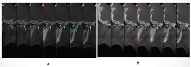
Figure 5: Sagittal computed tomogram showing no translation of the condyle. a: sagittal view of left condyle during maximal intercuspation position; b: sagittal view of left condyle during wide mouth opening position.
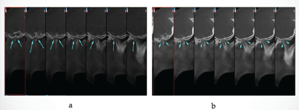
Figure 6: Sagittal computed tomogram showing no translation of the condyle, a: sagittal view of right condyle during maximal intercuspation position; b: sagittal view of right condyle during wide mouth opening position.
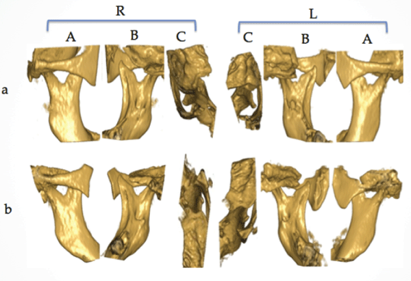
Figure 7: Cone beam computed tomography reconstruction showing the relationship between the coronoid process and the zygomatic arch. A: sagittal 3-D, lateral view; B: sagittal 3-D, lingual view; C: axial 3-D, upper view of the zygomatic arch; R: right, L: left; a: wide mouth opening position; b: maximal intercuspation position.
A 24-year-old female patient diagnosed at birth with AMC was referred to our faculty for limited mouth opening as the chief complaint. The medical history revealed that she had undergone surgical procedures, but she has no treatment regarding limited mouth opening. She seeks for the treatment of limited mouth opening, but she couldn’t be operated in her country. No contributory family and social history was reported.
The extraoral examination showed obvious deformities of the limbs, causing difficulty to take a step as in the first case. Alterations in the contraction pattern of the flexor muscles of the fingers were observed (Figure 8).
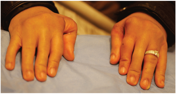
Figure 8: The contracted flexor muscles of the fingers.
There was no asymmetry of the face (Figure 9a). The translation of the condyles could not be detected, and there was no pain during the function and palpation of the masticatory muscles. During assisted opening, there was a hard end feel. The maximal mouth opening measured between the incisal edges of the maxillary and mandibular left incisors was 4 mm, and an absence of lateral and protrusive movements of the lower jaw was observed (Figure 9b). The intraoral examination showed no caries or periodontal problems. There were no malpositioned teeth within and between each arch (Figure 9c).
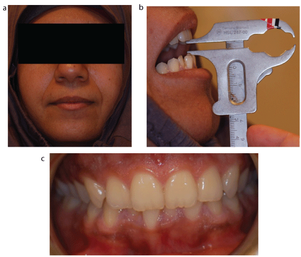
Figure 9: Clinical views of the patient. A: frontal view of the face; b: maximum mouth opening; c: frontal view of the occlusion.
Written informed consent was obtained.
Mandibular hypomobility results from many disorders affecting the stomatognathic system. The most common conditions related to this problem are those involving the temporomandibular joint and the masticatory muscles. Infrequently, congenital or developmental problems cause mandibular hypomobility, and arthrogryposis multiplex congenita is an example of these cases; it is characterized by multiple congenital contractures; in which joints in multiple body areas become limited by reduced fetal movement [14].
In the literature, the reports on AMC patients primarily focus on the limitation of limb movement and other physical conditions to improve the patients’ quality of life. Malnutrition, the negative effect on speech development, limited access to oral hygiene and dental care and muscle atrophy resulting from mandibular hypomobility in AMC patients should not be disregarded [23]. Twelve case reports on arthrogryposis multiplex congenita described mandibular hypomobility (PubMed, English language) [16,19-22].
In addition, affected patients frequently require anesthesia for the surgical release of contractures and correction of associated defects to enable function. For this reason, mandibular hypomobility in AMC patients is a matter of concern in anesthesia studies on difficult tracheal intubation during general anesthesia and in cases of limited neck extension and a short epiglottis [24,25].
In this case report, the first patient had speech problems, enlargement of the tongue, a severe open bite caused by mandibular hypomobility, a high maxillary palate, respiration problems and poor oral hygiene. The lateral and forward movement of the mandible was restricted because of obstruction of the enlarged coronoid process against the zygomatic arch. Although the patient had several surgeries for limb movement limitation, the family did not seek maxillofacial surgical treatment because of financial considerations, and the patient had adapted somewhat to the jaw movement limitation and managed to maintain compromised masticatory functions. She never had dental treatment until the occurrence of a toothache. According to clinical and radiographic evaluation, endodontic treatment was required for the left maxillary first molar diagnosed with deep dentin caries lesion. Limited mouth opening obstructed endodontic treatment, therefore the dental treatment involved an attempt of vital amputation.
In the second case, the patient didn’t have any dental problems. She seeked for the treatment of limited mouth opening. There was no micrognathia, cleft palate, high-arched palate, tongue with limited protrusion, dysphagia, weak masticatory and laryngeal muscles, poor gag and sucking reflex, microstomia, enlargement of the tongue, a severe open bite, respiration problems and poor oral hygiene as commonly seen in AMC patients.
Determining the cause of mandibular hypomobility in AMC patients is essential for establishing the appropriate treatment protocol. Mandibular hypomobility has been previously explained as being the result of muscle deficiency and tension arising from hypoplasia or atrophy [22]. Later in contrast to these hypotheses, the restriction of mouth opening was hypothesized to have a mechanical origin with an osseous contribution. Coronoid process hyperplasia is an infrequently encountered cause of limited mandibular jaw movement [3]. Although Iseberg et al. [26] has reported that coronoid hyperplasia is responsible for mandibular hypomobility in 5% of patients, the actual prevalence of coronoid hyperplasia is uncertain [27]. A review of literature reported limited number of cases of coronoid hyperplasia as the cause of limited jaw-opening movements in AMC patients in which there was no relevant TMJ or masticatory muscle morphological alteration [26]. The restriction of mouth opening in this case was of mechanical origin with an osseous contribution from the increased length of the coronoid process.
Various theories have been proposed for determining the cause of coronoid hyperplasia, including endocrine stimulation, increased temporalis activity, trauma, genetic inheritance and familial occurrence; however, the etiology of coronoid hyperplasia is unclear in AMC patients [4,23]. Coronoid hyperplasia might be related to calcification of the tendon of the temporalis [13]. Lack of joint movement also results in the tendons connected to the joint not stretching to their normal length, and short tendons case difficulty in normal joint movement [28]. The most accepted explanation relies on the hypothesis that reduced or absent mandibular movement and deglutition might lead to relative hyperactivity of the temporalis muscle that is not counterbalanced by the infrahyoid and suprahyoid muscles, thereby facilitating coronoid overgrowth [29]. In this case, there was no trauma during or after birth and no family history of AMC. The cause of restriction in these cases might be syndromic and associated with coronoid hyperplasia, which could cause a reduction in the height of the mandibular ramus.
A differential diagnosis of mandibular hypomobility is important to guide the treatment and improve the quality of life, and the use of appropriate imaging methods is essential for a definitive diagnosis [30]. The diagnosis is confirmed by panoramic radiographs and computed tomography scans. Evaluation of coronoid process enlargement could be performed by panoramic radiography, which is an easy, affordable and useful examination that provides a good overview of the mouth and adjacent tissues [31]. Computed tomography is an effective and efficient method for establishing the diagnosis. Three-dimensional cone beam computed tomography (3D-CBCT) provides a detailed anatomy of the coronoid process and the relationship with adjacent structures, revealing where impaction occurs in the zygomatic bone [3,32]. 3D-CBCT is significant in the diagnosis and surgical planning of coronoid hyperplasia [33]. In the cases presented, the clinical diagnosis of coronoid hyperplasia was verified using panoramic radiography and 3D-CBCT imaging techniques. The findings of the absence of bilateral condylar translation, the slender and pointed coronoid processes, the rudimentary condyles and the smooth condylar surfaces are similar to the findings of the previous studies.
There are a number of orthopedic articles focusing on the surgical treatment of joint involvement in AMC [16]. There is no specific treatment for AMC patients, and surgery is necessary to enable daily functioning [14]. Early manipulation soon after birth improves the passive and active range of motion.
Alignment and the establishment of stability for ambulation and functioning for self-care are principles from orthopedic surgery that can be applied to the maxillofacial region [14]. Treatment for limited mouth opening in AMC patients implies surgical correction of the mechanical interference by coronoidectomy and active physiotherapy [4,14]. Postoperative intensive physiotherapy following an adequate resection of the coronoid process is an accepted treatment to obtain satisfactory and stable results in the correction of coronoid-molar interference and for preventing postoperative fibrosis [34,35]. Difficulty in establishing a treatment protocol for AMC patients with mandibular hypomobility remains because of the limited number of patients with significantly limited range of motion [35]. Intraoral access to the coronoid process is the treatment of choice in 90% of the cases [14].
Although significant improvement is achieved by surgical treatment of coronoid hyperplasia, some authors have reported postoperative relapse because of fibrosis or coronoid regrowth [35-37]. Nordone et al. [14] stated that aggressive physiotherapy is important for improving joint motion and avoiding muscle atrophy and relapse.
In both cases, it is hypothesized that the mandibular hypomobility with AMC patients is caused by coronoid hyperplasia. The treatment option of coronoidectomy and physiotherapy was presented for both cases. The mother of the first case rejected the treatment for financial reasons. The second case also rejected the treatment because she lived in another country.
The two cases presented revealed that mandibular hypomobility in AMC patients is caused by coronoid hyperplasia, which is an uncommon reason that interferes with the patients’ quality of life. Three-dimensional computed tomography techniques are significant for obtaining an exact diagnosis and surgical planning for coronoid hyperplasia. Dentists should be aware of the rarely seen systemic conditions causing limited mouth opening.
BB: clinical examination, diagnosis, literature review, drafting and final approval of the manuscript; NG: diagnosis, literature review, drafting and Radiographic interpretation.
There is no funding for this article.
There are no conflicts of interest.
This case report does not require an Ethics Approval.
Download Provisional PDF Here
Article Type: Case Series
Citation: Bal B, Guler N (2017) Mandibular Hypomobility Caused by Coronoid Hyperplasia in Artrogryposis Multiplex Congenita: Two Case Reports and Literature Review. Int J Dent Oral Health 3(3): doi http://dx.doi.org/10.16966/2378-7090.232
Copyright: © 2017 Bal B, et al. This is an openaccess article distributed under the terms of the Creative Commons Attribution License, which permits unrestricted use, distribution, and reproduction in any medium, provided the original author and source are credited.
Publication history:
All Sci Forschen Journals are Open Access