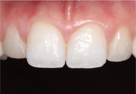
Figure 1: Initial clinical aspect of the case


Martini AP de Souza FI* Mazza LC da Cunha Melo RA Araújo NS Rocha EP
Department of Dental Materials and Prosthodontics, Araçatuba Dental School-Univ Estadual Paulista-Unesp, Brazil*Corresponding author: Dr. Fernando Isquierdo de Souza, Faculdade de Odontologia de Araçatuba-Unesp, Departamento de Materiais Odontológicos e Prótese, Rua José Bonifácio, 1193-Araçatuba-SP-16015-050, Brazil, E-mail: fernandofoa@hotmail.com
The treatment of conoids teeth represents a challenge for the dental team and, in particular, for the dentists who perform prosthetic restorations because only a few engagement elements are available in most cases, making it difficult to obtain an aesthetic that advocates anatomic and color homogeneity between the elements involved. Conoids teeth are changes in the size and shape of the natural teeth. These changes have an esthetic effect on the patient’s smile because the affected teeth are smaller than normal and have a tapered incisal surface. The most commonly used treatments are direct or indirect composite restorations with ceramic resin, with the former treatment being the preferred one. However, the wider use of ceramics has made it possible to obtain results that are more lasting and predictable because the cements provide adequate adhesion properties for the ceramic materials, which naturally mimic the aesthetics of the teeth; thus, these new materials have changed the aforementioned scenario. The correct treatment plan and the use of new adhesive ceramic materials has made the obtainment of good functional and esthetic results possible via an approach that is much more conservative than the composite resin treatment.
Conoids incisor; Laminate veneers; Dental ceramics; Lithium disilicate
Classified as an anomaly of size and shape of teeth, the conoid tooth are usually characterized by a size that is smaller than normal and a sharpened tip that replaces the nearly flat surface characteristic of the incisal edge of the maxillary lateral incisors. This anomaly manifests itself more frequently, affecting approximately 1% of the population, and is more common in females. It can also be associated with cases of agenesis or be a part of the characteristic signs of a syndrome [1-3].
This anomaly translates into esthetic discomfort for the patient, since the appearance of these teeth is far removed from the standards of normal. The most common treatment modalities include direct or indirect composite restorations ceramic resin, with the former treatment being the preferred one. However, this scenario has been transformed owing the widest possible use of ceramics in order to obtain a more predictable and durable treatment outcome [4].
With their excellent biomimetic characteristics, biocompatibility, durability, mechanical strength, and high chemical stability even in restorations with minimal thickness, ceramics are rapidly growing in restorative dentistry [5,6]. Depending on the needs of each case, a single ceramic system can meet the different demands, it restorations like crowns and veneers, contact lenses, or miniature ceramic fragments. Moreover, in this context, reinforced ceramic systems-alumina, zirconia, or especially in this case, lithium disilicate are able to provide increased mechanical resistance without impairing the esthetic results of restorations [7,8].
Furthermore, the realization of indirect restorations represents a major challenge to the dental technician, especially when referring to anterior teeth of young patients. The ability to reproduce specific technical aspects such as areas of translucency or the different shades and textures present in the adjacent teeth is what differentiates the outcome of this case, whose objective is to make the restoration in discernible when compared to the adjacent teeth.
When dealing with thin restorations, 1additional factor becomes critical to achieving success at the end of the restorative treatment: the capacity of the luting cement not to have a negative impact on the final color of the restoration, even over a long period [9]. There are kits available with a variety of luting cements for thin ceramic restorations. These kits are accompanied by shade guides, and ‘try-in’ enables the testing and selection of the most suitable color of cement to be used.
Thus, the reported clinical case meets certain interesting challenges to obtain an esthetic treatment outcome; for example, high esthetic expectations of the patient, young teeth characterized by predominant white patches resulting in a ‘milky’ appearance, and the necessity to make thin restorations that tend to some degree of transparency in order to respect the limits of the case.
The aim of this article is to describe a case in which thin veneers made of ceramic reinforced with lithium disilicate (IPS e.Max; Ivoclar Vivadent AG, Schaan, Liechtenstein) were used to achieve a satisfactory anatomy in maxillary lateral incisor conoids to meet the current esthetic standards without performing dental wear.
A 20-year-old female patient complained of poor esthetic appearance, both in relation to color and form of the direct composite resin restorations in teeth 12 and 22 (FDI system of tooth notation) that reanatomized the conoid teeth. Further, she reported uncertainty in performing everyday activities such as eating, for fear of fracturing the restorations.
On examination, it was found that both color and anatomy of the lateral incisors were unsatisfactory (Figure 1). Although there was dominance of the central incisors, the composition of the smile, in general, was satisfactory and constituted a harmonious whole, thus revealing the need for prosthetic intervention alone in the conoid teeth.

Figure 1: Initial clinical aspect of the case

The upper and lower dental arches of the patient were molded with addition silicone (Express XT; 3MESPE, St. Paul, MN, USA) and the casts were referred to the dental laboratory, for the models to be waxed.
Using the diagnostic wax-up, a mask was made with addition silicone (Express XT; 3MESPE, St. Paul, MN, USA) to prepare the mock-up for approval by the patient as well as the provisional restorations to be used while the preexisting restorations were removed. The treatment plan involved veneers for the lateral incisors using lithium disilicate ceramic (IPS e.max Press; Ivoclar Vivadent, Schaan, Liechtenstein).
The existing composite resin restorations were completely removed with the aid of sanding discs used for finishing resin restorations (SofLex Pop-on; 3M ESPE, St. Paul, MN, USA). No wear was performed on the dental tissues, with removal of only the pre-existing composite (Figure 2). Two retractor cords were inserted into the gingival crevice of the 2 teeth with the aid of appropriate instruments, with gauge in accordance with the gingival biotype of the patient (No. 00 and 0, Ultra pack; Ultra dent Products, South Jordan, UT, USA), and the impressions were made using addition silicone (Express XT; 3MESPE, St. Paul, MN,USA).
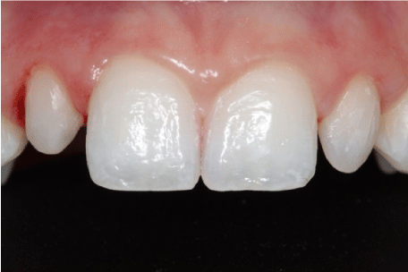
Figure 2: Clinical aspect after removal of composite resin restorations in teeth 12 and 22
The impressions were again sent to the dental laboratory for veneers to be fabricated. The color selection was performed with the aid of digital photographs and Vita Classical shade guide (VITA Zahnfabrik GmbH, Germany), as routinely performed by the laboratory (Figure 3).
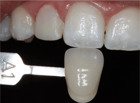
Figure 3: Photography for color selection of ceramics. Observe in the interim bisacrílica resin in the tooth 12
During the fabrication time of approximately 2 weeks for the ceramic restorations, the patient was provided with provisional restorations made of bis-acrylic composite resin (Protemp 4; 3MESPE, St. Paul, MN, USA). The provisional restorations has been poured into the mask obtained by molding the diagnostic waxing and positioned on the teeth of the patient after completion of the setting time of the resin, as indicated by the manufacturer (Figure 3).
The veneers received from the dental laboratory had thicknesses ranging from 0.4 mm to 2.0 mm in the incisal portion; these had been proven ‘draught proof ’, characterized by the simple placement of the restorations on teeth after removal of the provisional restorations. In this phase, we evaluated the adaptation of restorations to the dental substrate and the accuracy of the proximal contacts.
Further, there was a ‘damp proof ’ of ceramic restorations, which is made using shade guides of veneer cements that are specially developed for the cementation of thin parts. This phase is crucial for the color selection of the luting cement that will be used, so that it does not negatively affect the final color of the restoration, since the goal is the complete professional replication of natural teeth. After wetting with the try-in pastes, the proof of color ‘WO’ was selected for the cementation of restorations (RelyX Veneer, 3MESPE, St. Paul, MN, USA). Thereafter, we initiated the process of cementation of the restorations.
Ceramics enhanced with lithium disilicate are classified as acid sensitive and are therefore amenable to acid etching and silanization. The etching of the inner surface of the restoration was initiated using 10% hydrofluoric acid for 20s followed by washing with water and drying. Thereafter, application of 37% phosphoric acid for 1 min was followed by another round of washing with water and drying, and later, the application of silane (RelyX Ceramic primer; 3M ESPE, St. Paul, MN, USA). After waiting for the evaporation of the solvent as recommended by the manufacturer, the process ended with the application of the adhesive system (Single Bond 2; 3MESPE, St. Paul, MN, USA) and removal of the excess adhesive using an air jet for 20s [10].
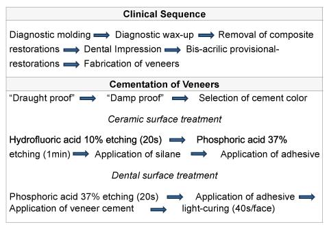
With the veneers ready for cementation, the teeth were etched using 37% phosphoric acid for 20s. After washing and drying the teeth, a layer of adhesive (Single Bond 2; 3MESPE, St. Paul, MN, USA) was applied and the excess adhesive was removed using compressed air for 10s, in relative isolation.
The cement RelyX Veneer (3MESPE, St. Paul, MN, USA) was applied directly to the inner surface of the restorations, and the restorations were correctly positioned on the teeth. After exposure to curing light (VALO, Ultra dent Products, South Jordan, UT, USA) to initiate polymerization for 5s on each side-buccal and palatal-the excess cement along the proximal surfaces was removed using an explorer and dental floss. Thereafter, each surface received curing light for 40s to complete the curing of the cement (Figure 4).
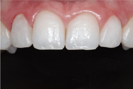
Figure 4: Immediate final aspect of the case
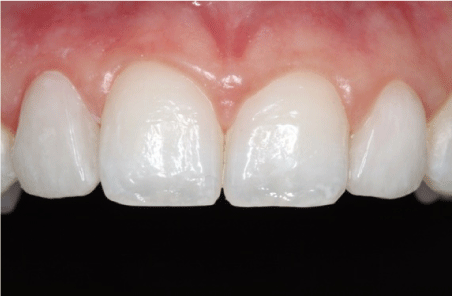
Figure 5: Aesthetic result after 2-year of cementation of restorations. Front view
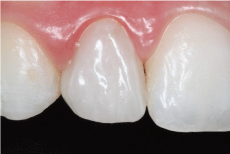
Figure 6: Aesthetic result after 2-year of cementation of restorations. Right Side View
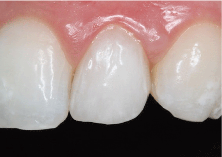
Figure 7: Aesthetic result after 2-year of cementation of restorations. Left side view
Figures 5-7 show, from different views, the esthetic treatment outcome achieved, after a 2-year follow-up period.
The search for a beautiful smile, or one that matches the esthetic standards of today, provides the growing use for ceramic restorations. The aim is to promote changes in the tooth form for example, the closure of diastemasor to promote color changes in pigmented teeth, besides replacing resin restorations that are direct or indirect. Such changes are likely to be carried out successfully owing to the characteristics of ceramics, which include greater clinical longevity, lower susceptibility to pigmentation, fewer fractures, and high biocompatibility characteristics that translate into satisfactory mechanical strength and high esthetics [11,12].
Survival rates of ceramic veneers in clinical studies have been reported to be 82.93% after 20 years in service, with 44.83% of the total failures caused by fractures in this period of observation [13], and in pieces cemented to the dentin substrate [12]. This high success rate is attributed, among other factors, to the conservation of tooth structure, because, the greater the thickness of the residual enamel, the higher the possibility of executing an effective bond with the ceramic [14].
Since the first report of the use of ceramic laminates in 1980, growing and dynamic advancements in ceramic and adhesive materials have enabled major advances in the use of such materials that reflect directly on the wear of the remaining tooth structure. Currently living in the reality of the concept of minimal invasion [15-17] and operating within its limits, this case report describes the fabrication of ceramic laminates in vital maxillary lateral incisor conoids without performing any tooth preparation.
In this context, the system reinforced with ceramic lithium disilicate that is fabricated by using a CAD/CAM processing manual or even pressing can be considered one of the best options to use, as in the presented case, since recent clinical studies [18] demonstrated very favorable structural integrity of restorations [19], with the absence of mechanical failures such as fracture or ‘chipping’, even in posterior teeth [20,21].
Considering the thickness of the ceramic restorations used in this case, it is noteworthy that changes in the color of the luting agent used may become visible at any time, thus negatively affecting the final esthetic outcome of the restorations [22]. The cement used in this clinical case as compared to other studies is fully photo-cured, indicating that these materials have high color stability, since they do not have amines in their composition. However, laboratory studies show a variation of opacity over time [23].
However, no clinical studies report long-term follow-up with appropriate means of evaluation, to verify the color stability of these anterior restorations in different intraoral conditions, which are indispensable to the needs of their current stage in dentistry, factor that constitutes a limitation for the evaluation of treatment performed. Nevertheless, long-term color stability is expected, because there were no changes of clinical or photographic conditions that might compromise the outcome of treatment on the period, and the patient remained satisfied. Finally, only after the completion of well-designed prospective clinical studies, can we answer accurately the critical question: to what extent do changes visualized in laboratory tests correspond, in reality, with the clinical performance of restorations?
This case report describes the anatomical correction of lateral incisor conoids with ceramic restorations after removal of the existing direct composite resin restorations, without performing any tooth preparation. The lithium disilicate-reinforced ceramic restorations seamlessly integrated with the adjacent teeth and caused no damage to the surrounding gingival during the 2-year follow-up period.
The authors have no financial interest in the products or companies mentioned in this study.
Download Provisional PDF Here
Article Type: Case Report
Citation: Martini AP, de Souza FI, Mazza LC, da Cunha Melo RA, Araújo NS, et al. (2016) Esthetic Treatment of Conoids Lateral Incisor Laminate veneers: A2-Year Follow-Up. Int J Dent Oral Health 2(4): doi http://dx.doi.org/10.16966/2378-7090.180
Copyright: © 2016 Martini AP, et al. This is an open-access article distributed under the terms of the Creative Commons Attribution License, which permits unrestricted use, distribution, and reproduction in any medium, provided the original author and source are credited.
Publication history:
All Sci Forschen Journals are Open Access