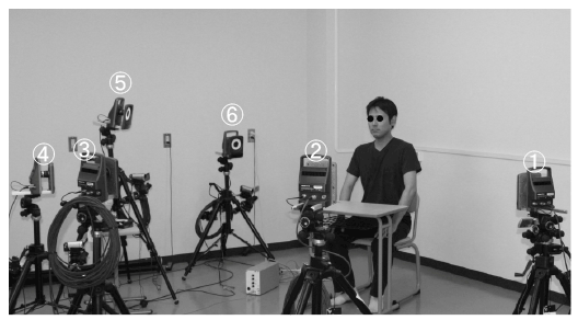
Figure 1: The motion-capture system. Numbers indicate each of the CCD cameras.

Ken Morizono1 Yoshihiko Takemoto*1 Emi Inada1 Daisuke Murakami1 Issei Saitoh2 Hideo Sato1 Tomonori Iwasaki1 Youichi Yamasaki1
1Department of Pediatric Dentistry, Kagoshima University Graduate School of Medical and Dental Sciences, 8-35-1 Sakuragaoka, Kagoshima 890-8544, Japan*Corresponding author: Yoshihiko Takemoto, D. D. S., Ph. D., Department of Pediatric Dentistry, Kagoshima University Graduate School of Medical and Dental Sciences, 8-35-1 Sakuragaoka, Kagoshima 890-8544, Japan, Tel: +81-99-275-6262; Fax: +81-99-275-6268; E-mail: yoshi-04@dent.kagoshima-u.ac.jp
Objectives: The purpose of this study was to evaluate the amount of lip movement and simultaneous tongue pressure changes on an artificial palatal plate during swallowing.
Methods: Subjects were 9 healthy males (25.4 ± 2.1 years of age). Three-dimensional lip movement was measured by a wireless optoelectronic system, and tongue pressure was simultaneously recorded by a sensor sheet attached to the incisive papilla of an artificial palatal plate. Reflective markers were attached to the right and left corners of the mouth to measure the distance between them. All subjects were instructed to swallow 5 mL and 20 mL samples of water at will. The maximum change of distance between the corners of mouth, the maximum tongue pressure, and the time interval between the two maxima (lip-tongue interval) were calculated. Wilcoxon’s test was used to detect significant differences in these measurements between the two volumes.
Results: Maximum tongue pressure did not differ significantly between swallowed volumes. The maximum change of distance between the corners of mouth was larger and the lip-tongue interval was significantly shorter with the larger volume.
Conclusions: We suggest that swallowing a larger volume is accomplished by larger lip movement rather than larger tongue movement. These results indicate that lip movement during swallowing can be evaluated objectively.
Swallowing; Lip motion; Tongue pressure; Motion capture; Simultaneous evaluation
Swallowing is a complex coordinated movement involving the craniocervical region, (i.e., neck, mouth, oropharynx and larynx) [1,2]. The characteristics of tongue, pharyngeal and laryngeal motion during swallowing are not easily evaluated from the body surface. In previous studies, swallowing was evaluated by observing subjects [3,4] and clinical patients [5] because it has been difficult to digitize the movement of the craniocervical region during swallowing in order to evaluate it objectively. Thus, tongue pressure [6-8], laryngeal elevation [9], frequency analysis of swallowing sounds [10] and so have been examined to quantitatively evaluate swallowing. Although these studies effectively digitized the movement of each organ during swallowing, each study evaluated only one organ. Evaluating more than two organs simultaneously could be a more effective method of evaluating swallowing, because swallowing is a complex coordinated movement of several craniocervical organs. Therefore, various methods (ultrasound imaging, video fluoroscopy, and video endoscopic examination) have been used to observe complex coordinated movement related to swallowing [11-14]. Although these methods can effectively evaluate swallowing movements, some of them can be harmful or inhibit natural movement [6]. People with dysphagia include growing children, the elderly, and some psychosomatically disabled, and they have a higher risk of aspiration and their swallowing is also more difficult to measure. So, a simpler and easier method of evaluating swallowing is needed.
Recently, the importance of lip form and pressure has been of interest to researchers and clinicians [15-17]. The lips are active when we make facial expressions, talk, breathe and eat, etc [18-20], and lip function may influence oral form [21-23]. Reduced eating and swallowing function because of lip dysfunction (due to a medical disorder or aging) are acknowledged as increasing the risk of aspiration [24]. So, methods to evaluate lip function during swallowing are needed.
Although there are no established methods for the assessment of lip function during swallowing, Ono et al. [25] suggested that measurement of lip pressure might be an effective method of evaluating swallowing. However, measurements made using a piece of material with a strain gauge between the lips could disturb natural movement of the lips during the swallowing.
The orbicularis oris is an important muscle involved in the oropharyngeal swallowing sequence, and also for lip closing, puckering the lips, and so on. Rosenblum considered activity of the modiolus, the conjunction of six facial muscles (the zygomaticus, caninus, triangularis, buccinator, platysma, and orbicularis oris muscles) at the lateral corners of the mouth to be useful for determining the manner of contraction of the orbicularis oris [3]. Moreover using physiographic cinematography, he confirmed that the occurrences of modiolus activity during swallowing are involved in extending the corners of the mouth from the anatomical position [3]. Although the lips have been shown to perform an important function during normal swallowing by various measurements, there have been very few quantitative evaluations of the association between lip movement and swallowing. Therefore, we decided to examine the movement of the corners of the mouth near the modioli as an index of lip movement in this study.
Accordingly, the purpose of this study was to quantitatively evaluate the movement of the lips during swallowing with a simple, easy and noninvasive method from the body surface and simultaneous tongue-pressure changes, which have been used in past studies to evaluate swallowing [6-8].
Nine men (average age, 25.4 ± 2.1 years) participated in this study. None of the subjects experienced pain or dysfunction in the craniomandibular region, and none had tooth loss except for third molars, abnormalities of occlusion or history of dysphagia. Informed consent to participate in this study was obtained from all subjects. Prior to beginning the study, approval of the clinical ethics committee of Kagoshima University Hospital had been obtained (No.26-112).
The Vicon system® (Inter-Reha Co. Ltd., Tokyo, Japan) was used as an optical 3-D motion analyzer. Light-reflective balls were attached to the skin of each subject, and these markers’ motion was tracked with six charge-coupled device (CCD) cameras and visualized on a computer display. Six CCD cameras were placed one meter away from each subject (Figure 1). The markers’ three-dimensional coordinates were tabulated at 100 Hz.
Retro-reflective markers (diameter, 3.5 mm) were attached to the right and left corners of the mouth to record lip movement during swallowing (Figure 2). Because the markers were small and lightweight the subjects could swallow water as usual.
Before measuring tongue pressure, all subjects had maxillary toothform impressions taken for making a 0.75 mm thick artificial palatal plate (IMPRELON® S, Scheu-Dental, Iserlohn, Germany). A small-sized pressure sensor (PSS Subminiature Pressure Sensor®, Kyowa Electronic Instruments, Tokyo, Japan) was attached to the thickest part of the incisive papilla on the artificial palatal plate with soft resin as described by Hori et al. [6]. In order to limit the amount of subjects’ occlusal height change, the path of the sensor cable was designed to go around the last upper molar tooth and then out of their mouth (Figure 3). During the measurement of tongue pressure, the sensor sheet was tightly fitted to the palate and the subject could swallow water with minimum discomfort because of its thinness. After measurement, the palatal plate was unstuck safely.
Figure 1: The motion-capture system. Numbers indicate each of the CCD cameras.
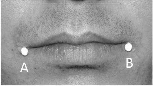
Figure 2: Anterior view of a mouth of subject showing the location of the 3.5 mm retro-reflective markers on the right (A) and left (B) corners of the mouth.
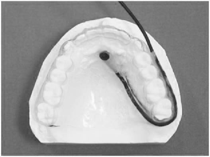
Figure 3: The pressure sensor attached to the considerable part of incisive papilla on the artificial palatal plate, and the path of the sensor cable.
Before beginning the swallowing measurements the resting condition was recorded for five seconds while subjects kept their mouth closed in a natural head position, and the “Resting distance” between the corners of mouth was calculated. Next, subjects were instructed to sit upright with a natural head position. The subjects were instructed to swallow a mouthful at a self-decided time. Prior to recording, either a 5 mL or a 20 mL water sample was then carried to the mouth of each subject [26], and recording began. After a signal to start, subjects voluntarily swallowed the water when ready. Recording ended after subjects raised their hand after having swallowed the water. Lip motion and tongue pressure during swallowing were simultaneously recorded. Subjects repeated swallowing each volume three times, for a total of six trials. The subjects had a 1-min rest between recordings.
The three-dimensional coordinates of each marker’s position relative to the floor and the tongue pressure were simultaneously recorded at 100 samples/sec.
Maximum tongue pressure (MTP): The maximum magnitude of tongue pressure (MTP) was determined by plotting the pressure over time and identifying the peak value of the vertical axis of the waveform (Figure 4). Because Hori et al. [6] and Furuya et al. [7] indicated that tongue pressure is an effective method for evaluating swallowing; MTP was considered as the index of tongue elevation during swallowing in this study. The unit of MTP was kPa.
Maximum lip motion (MLM): The distance between the two markers of the right and left corners of the mouth during the five seconds of resting condition (Resting distance) and during swallowing (Swallowing distance) were calculated from the three-dimensional coordinates of each marker. The maximum magnitude of lip movement (MLM) was determined by plotting the distance between the corners of the mouth relative to the resting distance and identifying the peak value of the vertical axis of the waveform (Figure 4). MLM was considered as the index of lip motion, and the units of MLM are mm.
The interval between lip and tongue actions (L-T interval): To examine the time interval between lip and tongue action during swallowing, the L-T interval was calculated according to the formula:
L-T interval = T(MTP) - T(MLM) [msec]
Where T(MLM) and T(MTP) represent the time of MLM and the time of MTP, respectively. A graphic example of this calculation is shown in Figure 4.
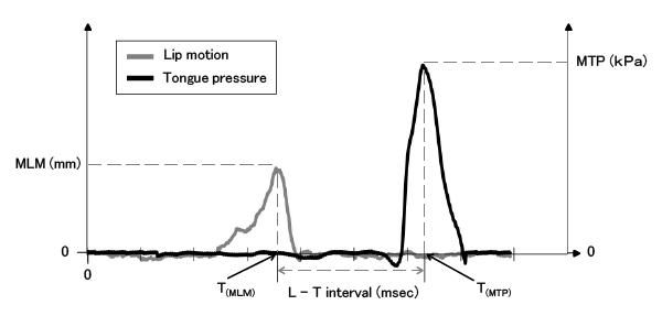
Figure 4: Representative waves of tongue pressure and lip motion, and items for evaluating the L-T interval during 5 mL water swallow.
After the three measurements for each volume were averaged for each subject, Wilcoxon signed-rank test was used to detect significant differences between swallowing of the two volumes of water. Multilevel linear model analysis was used to evaluate MTP, MLM and L-T interval for each phase and the variances at both the inter-individual and intraindividual levels using MLwiN® software (University of Bristol, version 2.0). Multilevel models estimate variance based upon the results of an iterative generalized least-squares method. Each multilevel model is composed of two parts, fixed and random. The fixed part estimates the population parameters, which closely correspond to the mean estimates of traditional analyses, and the standard errors associated with each parameter are also estimated. The random part estimates variation at different hierarchical levels, with one level nested within the preceding level. A two-level model was used to estimate the mean and standard error of the mean for significance [27]. The two levels pertained to random variation of MTP, MLM and L-T interval between the participants (interindividual) and between repeated measurements (intra-individual).
MTP did not differ significantly between the 5 mL and 20 mL volumes of water (Figure 5A). The magnitude of MLM was significantly larger, and the L-T interval was significantly shorter when swallowing the larger volume. (Figures 5B and 5C).
Inter- and intra-individual variation of MLM, MTP, and the L-T interval are shown in Table 1. Intra-individual variation of all variables was smaller than the inter-individual variation. Both the inter- and intra-individual variations of MTP when swallowing 5 mL of water were smaller than when swallowing 20 mL of water. On the other hand, both the inter- and intra-individual variations of MLM and the L-T interval when swallowing 5 mL of water were larger than when swallowing 20 mL of water. We show an example of the MTP, MLM and L-T interval when a subject drinks the different amounts of water in Figure 6.
A swallow has four phases: oral preparatory, oral propulsive, pharyngeal and esophageal. Swallowing from the oral propulsive phase to the pharyngeal phase is a complex act that involves the coordinated activity of several organs, including the lips, organs during swallowing. Hori et al. [28] reported that tongue pressure produced bolus tongue, pharynx, and larynx. Previous reports examined the relationships among this propulsion and temporally were closely related to hyoid movement during swallowing. Ertekin et al. [29] showed that the orbicularis oculi and orbicularis oris muscles were synchronously activated during spontaneous swallowing in humans.
Furthermore, many studies have also reported that head position can influence the process of swallowing. Woo et al. [30] tested the effects of cranio-cervical flexion (CCF) on activation of the swallowing-related suprahyoid and infrahyoid muscle group (which is located in the same area as the superficial neck flexor muscles) while swallowing liquids. They suggested that changing CCF might be a viable method to enhance the effectiveness of the swallowing-related muscles.
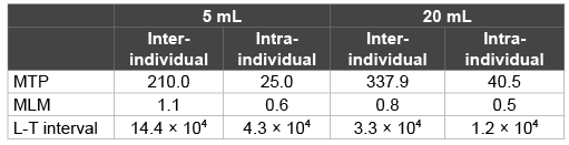
Table 1: The inter- and intra-individual variations of MTP, MLM, and L-T interval
Although these reports have established the importance of coordinated movement in the craniocervical region during swallowing, the normal sequence of events during swallowing remains unknown. Furthermore, the importance of lip movement from the oral propulsive phase to the pharyngeal phase of swallowing is not well understood. Tamura et al. [31] compared lip function during swallowing between senile subjects with and without a history of aspiration, and reported weaker vertical labial pressure and less lip movement in subjects who experienced “choking on food”. Their results suggest that vertical labial pressure and lip movement play an important role in swallowing.
Additionally, Ono et al. [25] indicated that lip closure is highly important with regard to ingestion of food into the oral cavity, mastication, and swallowing of the bolus.
Although lip pressure and lip-closing force have been used for the evaluation of lip function in swallowing in past studies [6,25,31], the methods they used may have disturbed spontaneous lip movement. When lip movement during swallowing can be evaluated quantitatively from the body surface, spontaneous lip function is less likely to be disrupted. Therefore, we reason that MLM can be used to evaluate spontaneous lip function during swallowing. Rosenblum et al. [3] stated that activity of the modiolus, can be observed during swallowing in many subjects by using physiographic cinematography, and that an increase in inter-modiolus distance is associated with extension of the corners of the mouth. In this study the distance between the corners of the mouth during swallowing increased in all subjects. Therefore, we suggest that the muscular action around the lips, as indicated by modiolus movement (MLM), increases when the quantity of water increases.
In contrast, Hori et al. [32] found no difference in peak magnitude of tongue pressure among bolus types, the same as our results for MTP. Tongue elevation in normal subjects is thought to be a relatively stable action regardless of the bolus volume. Accordingly, past studies and our results suggest that during swallowing of an increased amount of water lip movement compensates because tongue elevation remains stable (Figures 5A and 5B).
Lip action tends to begin earlier than tongue action in normal adults’ swallowing, as indicated by the L-T interval. This observation was possible only because in this study lip movement and tongue elevation were evaluated simultaneously. In addition, the shorter lip-tongue interval with the larger volume suggests that adjustment of lip movement is associated with controlling the timing of swallowing (Figure 5C).
Smaller intra-individual variation of MTP and MLM than interindividual variation showed that individuals have stable patterns of tongue and lip function. Inter- and intra-individual variation of MLM decreased with the increased volume of swallowed water, while the reverse was true for MTP. The smaller inter- and intra-individual variation in the L-T interval with 20 mL of water than 5 mL suggests limited swallowingpattern flexibility, because swallowing the larger volume required greater coordination between the tongue and lips. Ertekin et al. reported that 20 mL of water was the upper limit of the quantity that normal subjects could swallow by a mouthful [26]. Therefore, we suggest that more closely coordinated movement between the lips and tongue are needed when swallowing 20 mL than 5 mL.
In summary, simultaneous recording of lip movement and tongue pressure and displaying the coordinated movement of the lips and tongue show that tongue pressure is stable and lip motion changes more with changes of the amount of swallowed water (Figure 6). Our results indicate that normal individuals exhibit a consistent pattern of swallowing, and simultaneous evaluation of two organs effectively demonstrated changes of coordinated action between the two organs during swallowing.
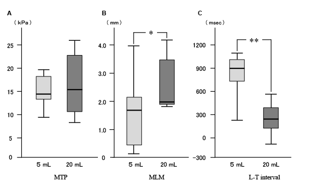
Figure 5: Comparisons of the MTP (A), MLM (B), and L-T interval (C) for 5 mL water swallowing and 20 mL water swallowing. Solid bars within each box indicate medians, * and ** indicate significant differences at p<0.05 and p<0.01.
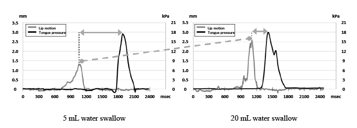
Figure 6: Tongue pressure and lip motion waves from one subject showing 5 mL water swallow and 20 mL water swallow. The waves show clearly that MLM was larger, and L-T interval was shorter in swallowing the larger volume.
Lip movement during swallowing was objectively evaluated, and we concluded:
This work was supported by KAKENHI from Japan society for the Promotion of Science (No. 15K20604)
Download Provisional PDF Here
Article Type: Research Article
Citation: Morizono K, Takemoto Y, Inada E, Murakami D, Saitoh I, et al. (2016) Simultaneous Evaluation of Three-Dimensional Lip Kinetics and Tongue Pressure during Swallowing. Int J Dent Oral Health 2(2): doi http://dx.doi.org/10.16966/2378-7090.169
Copyright: © 2016 Morizono K, et al. This is an open-access article distributed under the terms of the Creative Commons Attribution License, which permits unrestricted use, distribution, and reproduction in any medium, provided the original author and source are credited.
Publication history:
All Sci Forschen Journals are Open Access