Figure 1: Initial intraoral photographs: A – right side; B – frontal and C – left side.

Andre Wilson Machado* Emanuel Braga
1Associate Professor, Section of Orthodontics, Federal University of Bahia, Dental School, Salvador / BA, Brazil*Corresponding author: Andre Wilson Machado, Av. Araújo Pinho, 62, 7º andar, Federal University of Bahia, School of Dentistry, Salvador/BA, Brazil, Tel: 55 [71] 3334-1163; E-mail: awmachado@bol.com.br
A mesiodens is a supernumerary tooth located in the maxillary central incisor region and may be a cause of malocclusion in this region. We report an uncommon severe sequelae after an unerupted mesiodens removal in the permanent dentition related to midline bone loss, gingival recession and loss of vitality of the left upper central and lateral incisors. This article is written to highlight that in order to avoid unwanted collateral effects, surgical removal of mesiodens must be indicated only in specific conditions.
Mesiodens; Supernumerary teeth; Complications; Maxillofacial surgery
Supernumerary teeth are teeth in excess of the normal number [1]. When located in the maxillary central incisor region a supernumerary tooth is called mesiodens and its prevalence has been estimated to be 0,15 to 2,2% of the population with a preference of male [2]. Mesiodens is usually found to be impacted but also fully or ectopic erupted and only 6% are found in a horizontal position [3].
The etiology of mesiodens remains unknown, although different theories are formulated. Among those, the most accepted states that locally induced hyperactivity of the dental lamina, influenced by environmental and genetic factors, results in supernumerary teeth. It is also important to remember that due to its familial occurrence, mesiodens etiology has a strong correlation with genetics [2-4].
It is important that the diagnosis of mesiodens should be made as early as possible due to its associating with disturbances in tooth eruption such as delayed eruption of the permanent incisors, crowding or interference with the alignment of the maxillary incisors, dental impaction, resorption of adjacent tooth, development of dentigerous cyst, facial sinus and one of the most common sequel, a midline diastema [3-5].
Treatment options for mesiodens may include surgical extraction or monitoring without removal [4,6]. Although mesiodens extraction may seem to be a simple procedure its surgical approach could jeopardize the development of adjacent teeth as well as the surroundings tissues. Therefore, this paper presents an uncommon severe sequelae after an unerupted mesiodens removal in the permanent dentition related to midline bone loss, gingival recession and loss of vitality of the left upper central and lateral incisors.
A 30-year-old female Brazilian patient was referred to the first author private office for orthodontic evaluation. Her medical and dental histories were uneventful.
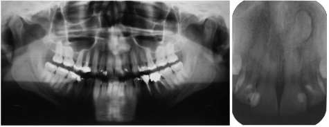
The patient exhibited a symmetric face with characteristics within normal standards and a close-up assessment revealed a pleasant smile with good exposure of upper incisors. Intra-orally there was a Class I malocclusion with good upper incisors alignment and a healthy perioduntium (Figure 1). Radiographic examination revealed the presence of an asymptomatic horizontal mesiodens. The use of a tube shift technique indicated that it was positioned palatally (Figure 2).
Orthodontic planning was designed to obtain the correct alignment, leveling and dental intercuspation as well as to extract the lower left first molar and closing the remaining space with forward movement of the second and third molar. Before the orthodontic treatment was initiated patient was referred to an oral maxillofacial surgeon for the mesiodens removal.
The surgical procedure was carried out under local anesthesia and initially, mesiodens was approached palatally. Though, during surgery procedure due to the mesiodens close proximity to the adjacent incisors roots a labially intervention was also necessary.
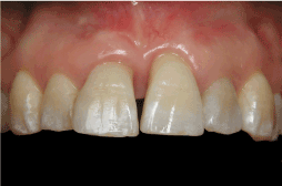
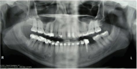
After surgery, the patient developed an important gingival recession in the upper left central incisor associated with bone loss in the mesial surface (Figure 3). Four months after surgery, pulp vitality tests of the maxillary anterior upper teeth confirmed the loss of vitality of the left central and lateral incisors and thus, root canal therapy were indicated (Figure 4).

Figure 1: Initial intraoral photographs: A – right side; B – frontal and C – left side.

Figure 2: Initial radiographs: A – panoramic and B – periapical.

Figure 3: Intraoral close-up photography one month after surgery.

Figure 4: Panoramic radiography 4 months after surgery.
It is strongly advised that the diagnosis of mesiodens should be made at an early phase possible due to its associating with disturbances in tooth eruption [3-5]. This is not the case in this clinical report since the patient was referred to the orthodontist at 30-year-old with an undiagnosed and asymptomatic that seemed to be innocuous to the occlusion. It is therefore important for the clinician to diagnose a mesiodens early in development to allow for optimal yet minimal treatment [4].
Treatment options for mesiodens include surgical extraction or monitoring without removal [4,6]. Ultimately, the question is whether to leave the mesiodens alone or remove it. Surgical removal must be considered in specific conditions such as: [a] if the supernumerary presence poses a risk of damaging the developing surrounding teeth; [b] there is associated pathology or [c] if it presents an obstacle to planned orthodontic tooth movements [7]. The latter condition was the main reason the patient was referred for mesiodens removal since orthodontic movement could damage the maxillary incisor roots if they came to contact with the mesiodens. Conversely, if the mesiodens remains in place without symptoms and does not adversely affect the adjacent teeth, it may be left in place and observed periodically and thus, surgical removal must be contraindicated [7].
In the case presented, midline bone loss, gingival recession and pulp necrosis were postoperative complications of the surgery. Although the explanation of these sequelae after a mesiodens removal is not very well documented in the literature, understanding the complications followed by orthognathic surgery could be extrapolated to clarify those side effects [8].
Some studies have demonstrated that successful oral and maxillofacial surgery requires maintenance of an adequate blood supply and that vascular integrity is a critically important factor in preserving the skeletal component and also the dentition and the associated soft tissue elements [pulp, periodontium, and gingival] [8]. Studies on humans have also demonstrated that long-term ischemia in orthognathic surgery could lead to the development of periodontal defects and pulp degenerative changes [9]. Regarding this issue, Wolford [10] has pointed out that since the blood supply to the anterior segments is primarily through the palatal mucoperiosteum any trauma in this area can cause major vascular risk to bone, teeth, and soft tissues, creating significant periodontal problems and possible loss of these structures. Considering this background, the authors hypothesized that although the postoperative complications found in this case report are interrelated, it could be associated to some major aspects such as: [a] the surgery trauma itself; [b] a palatally decreased blood flow associated with the surgery; [c] the significant amount of bone removal; [d] the mesiodens proximity to the incisors root apex. This latter aspect highlights the need of computed tomography [CT] evaluation before surgery to have a more accurate position of the mesiodens and the surrounding structures [1]. For example, Becker et al. [11] demonstrated that inaccurate 3-dimensional diagnosis of impacted canines was proven to be one of the major reasons for treatment failure. In the case reported, only conventional radiographs [panoramic and periapical] were assessed which definitely had contributed to increase the risk of intraoperative complications.
It is important to remember that although those complications were reported in the orthognathic surgery literature [12] there is still lack of evidence that a mesiodens surgery removal, which is a much more simple procedure than the orthognathic procedure could lead those complications.
The analysis of the results after surgery showed that not only the smile aspect was deteriorated but also the patient reported a lower level of selfconfidence and was generally unhappy with the surgery result.
In summary, it is of paramount importance to point out that although mesiodens removal is suggested in the literature, surgical procedure should be indicated only if the supernumerary jeopardizes the normal development of the dentition or even if it imposes a risk to the orthodontic treatment. In the case described the surgery had a deleterious impact in the functional and esthetic standpoint and thus, although mesiodens surgery removal has proven to be safe, patients who undergo such surgery must be aware of all types of side effects including the one related in this case report.
Download Provisional PDF Here
Article Type: Case Report
Citation: Machado AW, Braga E (2015) Severe Sequelae After A Mesiodens Surgical Removal: A Clinical Report. Int J Dent Oral Health 1(4): doi http:// dx.doi.org/10.16966/2378-7090.124
Copyright:© 2015, Machado AW, et al. This is an open-access article distributed under the terms of the Creative Commons Attribution License, which permits unrestricted use, distribution, and reproduction in any medium, provided the original author and source are credited.
Publication history:
All Sci Forschen Journals are Open Access