
Full Text
Effect of Nanofiller Technology on Surface Properties of Nanofilled and Nanohybrid Composites
Neha Jain1*, Arti Wadkar21Reader, Department of Prosthodontics, Sudha Rustagi College of Dental Sciences and Research, Faridabad, Haryana, India
2*Professor, Department of Prosthodontics, Nair Hospital and Dental College, Mumbai, India
*Corresponding author: Dr. Neha Jain, Flat no. 403, Tower no. 15, The Close North Apartments, Sector- 50, Gurgaon, India, Tel: 09818095343; E-mail: nejamaverick@gmail.com
Abstract
Context: Nanocomposites are becoming popular today because of the manufacturers claiming high surface smoothness and good abrasion resistance properties thus improving the longevity of the restoration.
Aims: This in vitro study was carried out to investigate the effect of nanofillers on surface roughness and abrasion resistance of nanofilled composite- Filtek Z350 (3M ESPE) and nanohybrid composite- Tetric N Ceram (Ivoclar Vivadent) after polishing and abrasion.
Methods and material: A total of fifteen disc specimens of each category of material were prepared under standardized conditions. The discs were finished and polished according to manufacturer’s instructions. The samples were examined for surface roughness using Mahr Perthometer M2 followed by scanning electron microscope examination. In order to assess the performance of the materials in simulated oral conditions the samples were then subjected to toothbrush-dentifrice abrasion and the resultant surface roughness was compared for both the materials.
Statistical analysis: Mean value of surface roughness was calculated for both the groups from Ra values before and after toothbrush-dentifrice abrasion followed by calculation of Standard deviation (SD). Paired-t test was then applied to compare the surface roughness values before and after toothbrush-dentifrice abrasion within the same group.
Results: Initially the surface roughness of Filtek Z350 were found to be superior to Tetric N Ceram but after subjecting to toothbrush dentifrice abrasion Tetric N Ceram showed greater increase in surface roughness as compared to Filtek Z350. The difference was found to be statistically significant.
Conclusions: Nanofiller type, size and distribution significantly influence the surface properties of composites. Though nanohybrid composite showed a better initial surface polish but nanofilled composite showed a better abrasion resistance.
Keywords
Nanocomposites; Nanofillers; Surface properties; Dental restoration wear
Introduction
Filler particle size and morphology are very crucial to the physical properties and clinical performance of composites [1]. Microfilled composites were the first materials to be sufficiently wear resistant and maintain an acceptable surface quality due to small filler particles and low filler content. However, microfilled composites face two major disadvantages viz high polymerization shrinkage and low flexural strength. Hybrid composites on the other hand possess fillers of different sizes leading to high filler content and hence show higher physical strength and acceptable polymerization shrinkage. However they exhibit poor surface polish retention [2-6].
Nanocomposites have been recently introduced to serve these functional needs through the application of nanotechnology [7]. They have improved mechanical properties i.e. better compressive strength, diametrical tensile strength, fracture resistance, wear resistance, low polymerization shrinkage, high translucency, high polish retention and better esthetics [8,9]. With such excellent properties they could turn out to be a cost-effective, time saving and easy to repair and finish alternative to ceramics as laminate materials.
Nanotechnology has been used in composites in the form of nanofilled and nanohybrid composites which possess different filler morphology, particle size and distribution. This study was undertaken to assess the effect of this difference on two main surface properties viz surface roughness and abrasion resistance of nanofilled (Filtek Z350, 3M ESPE) and nanohybrid composite (Tetric N Ceram, Ivoclar Vivadent).
Materials and Methods
Preparation of samples
A custom made brass mould consisting of holes 8 mm in diameter and 0.8 mm in depth was used to fabricate 15 disc samples each of Filtek Z350 (3M ESPE) and Tetric N Ceram (Ivoclar vivadent). Samples were divided into two groups according to the material: a) Group 1- Filtek Z350, b) Group 2- Tetric N Ceram. The details of the materials are mentioned in Table 1.
| Grp no. | Composite | Type | Lot no. | Manufacturer | Composition | Filler loading (wt%) |
|---|---|---|---|---|---|---|
| 1 | Filtek Z350 | Nanofilled | 20070628 | 3M ESPE, USA | Matrix: Bis-GMA, UDMA,
TEGDMA, and Bis-EMA resins
Filler: a) Non-agglomerated 20 nm silica filler
b) Non-agglomerated 4 to 11 nm zirconia filler c) Aggregated zirconia/silica cluster filler (comprised of 20 nm silica and 4 to 11 nm zirconia particles) in the size range of 0.6 to 1.4 microns |
78.5 |
| 2 | Tetric N Ceram | Nanohybrid | K04764 | Ivoclar vivadent, Schaan | Matrix: Bis-GMA, UDMA, TEGDMA, Bis-EMA resins |
80.5 |
Table 1: Systemic diseases chosen for the study along with the most frequent forms of the disorders seen in the sample.
|
Mean |
N |
Std. Deviation |
Std. Error Mean |
|
Group -1 |
Average surface roughness before toothbrush- dentifrice abrasion |
0.367 |
15 |
0.037 |
0.009 |
Average surface roughness after toothbrush- dentifrice abrasion |
0.544 |
15 |
0.029 |
0.007 |
|
Table 2: Systemic diseases chosen for the study along with the most frequent forms of the disorders seen in the sample.
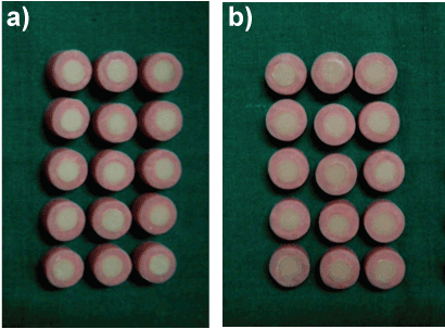
The material was dispensed from the syringe and packed in the mould placed on a glass slab and covered with Mylar strip. The excess was removed and the sample was cured with L.E.D. light cure unit (Optilight LD Max, Gnatus, Voltage: 93 V–260 V, Frequency: 50/60 Hz, Power: 15 VA, Wave length: 440 nm-460 nm) for 40 seconds. Custom made metal mould of dimensions 10 mm in diameter and 10 mm in height was used for fabrication of autopolymerizing acrylic resin blocks (DPI-RR - pink, India, working time of 4 min and bench cure time of 14 min) to embed composite disc samples keeping one side of the disc exposed (Figures 1a and 1b). Samples were then subjected to their polishing systems i.e. Astropol polishing kit and Sof-lex polishing kit for Tetric N Ceram and Filtek Z350 respectively as recommended by the manufacturers.
Testing the surface roughness
All the specimens were tested for surface roughness using Surface Roughness Tester-Mahr Perthometer M2, MAHR GMBH, Germany (Figure 2). It measures range of up to 150 μm/6000 μin, and traverses lengths 1.75 mm, 5.6 mm, 17.5 mm (.07 in, .22 in, .7in) as per ISO Standards. The values were obtained by means of a stylus instrument with a 2 μm tip and a cone angle of 90° (as per DIN EN ISO 3274) which enabled a two-dimensional tracing of the surface. Five successive measurements in different directions were recorded for each specimen and the average surface roughness (Ra, μm; as per DIN EN ISO 4287) was calculated. Representative samples of each group were examined under scanning electron microscope (FEI Quanta 200 Mark 2) and photomicrographs were taken (Figures 3a and 4a).
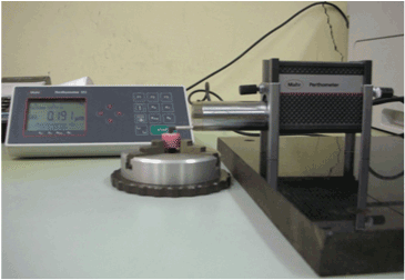
Figure 2: Measurement of surface roughness using Mahr’s Perthometer
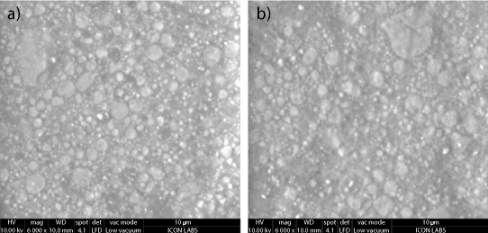
Figure 3: Photomicrographs of Filtek Z350 a) Before toothbrushdentifrice abrasion nanoclusters and nanofillers surrounded by resin matrix b) After toothbrush-dentifrice abrasion loss of nanofillers but nanoclusters stay intact.
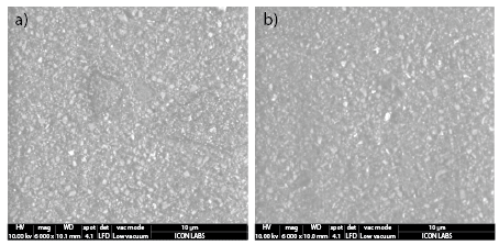
Figure 4: Photomicrographs of Tetric N Ceram a) Before toothbrushdentifrice abrasion different filler particles surrounded by resin matrix b) After toothbrush- dentifrice abrasion loss of large size filler particles.
Toothbrush-dentifrice abrasion
Specimens were then subjected to toothbrush-dentifrice abrasion with a powered toothbrush (Oral B Cross Action with round end, medium bristles and speed of 7200 rpm) and dentifrice (Pepsodent toothpaste, Hindustan Unilever Ltd, India, containing calcium carbonate) followed by testing for surface roughness with Mahr Perthometer M2. A custom made jig was used to hold the brush head perpendicular to the disc sample. A brushing sequence of 36,000 cycles was performed under a constant load of 500 gm (applied on the brush head) for 5 min in slurry of distilled water and dentifrice in the ratio of 1:1 by weight [10-12]. Average surface roughness (Ra, μm) for each specimen was again calculated post toothbrush dentifrice abrasion. This was followed by examination under scanning electron microscope and photomicrographs were taken (Figures 3b and 4b).
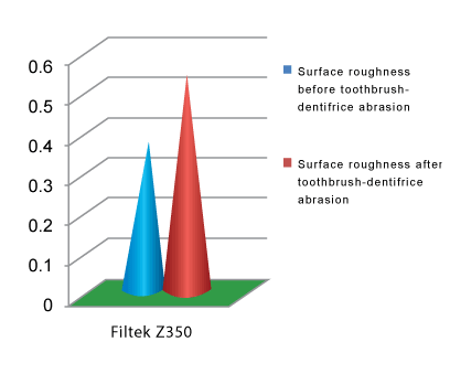
Graph 1: Mean surface roughness values before and after toothbrushdentifrice abrasion for Group I
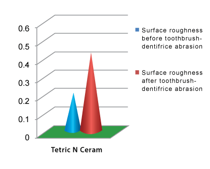
Graph 2: Mean surface roughness values before and after toothbrushdentifrice abrasion for Group II
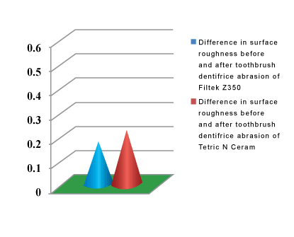
Graph 3: Difference in surface roughness values of Groups I and II before and after toothbrush-dentifrice abrasion
Statistical analysis
Mean value of surface roughness was calculated for both the groups from Ra values before and after toothbrush-dentifrice abrasion followed by calculation of Standard deviation (SD). Paired t test was then applied to compare the surface roughness values before and after toothbrushdentifrice abrasion within the same group.
Results
As seen in Tables 2 and 4 surface roughness of Group-1 was 0.367 ± 0.037 μm and Group-2 was 0.210 ± 0.015 μm before toothbrush-dentifrice abrasion. This indicated that surface roughness of Tetric N Ceram was lower than that of Filtek Z350 and the difference was found to be statistically significant. Surface roughness values increased after toothbrush-dentifrice abrasion. Surface roughness of Groups 1 and 2 was 0.544 ± 0.029 μm and 0.430 ± 0.028 μm respectively. Hence surface roughness of Tetric N Ceram was still lower than Filtek Z350 and the difference was statistically significant. Paired-t test results (Tables 3 and 5) showed that the paired difference in the values was statistically significant for both Filtek Z350 and Tetric N Ceram at 0.05 level of significance. Comparison of the difference of the mean values of both the groups before and after toothbrushdentifrice abrasion as seen in Table 6 showed that the difference was more for Tetric N Ceram as compared to Filtek Z 350.
|
Mean |
Std. Deviation |
Std. Error Mean |
t |
df |
Sig. (2-tailed) |
|
Group 1 |
Average surface roughness before toothbrush- dentifrice abrasion - Average surface roughness after toothbrush- dentifrice abrasion |
-.176480 |
0.037481 |
0.009678 |
-18.236 |
14 |
<0.0001 |
Table 3: Systemic diseases chosen for the study along with the most frequent forms of the disorders seen in the sample.
|
Mean |
N |
Std. Deviation |
Std. Error Mean |
|
Group 2 |
Average surface roughness before toothbrush- dentifrice abrasion |
0.210 |
15 |
0.015 |
0.004 |
Average surface roughness after toothbrush- dentifrice abrasion |
0.430 |
15 |
0.028 |
0.007 |
|
Table 4: Systemic diseases chosen for the study along with the most frequent forms of the disorders seen in the sample.
|
Mean |
Std. Deviation |
Std. Error Mean |
t |
df |
Sig. (2-tailed) |
|
Group 2 |
Average surface roughness before toothbrush- dentifrice abrasion - Average surface roughness after toothbrush- dentifrice abrasion |
-0.219627 |
0.025851 |
0.006675 |
-32.905 |
14 |
<0.0001 |
Table 5: Systemic diseases chosen for the study along with the most frequent forms of the disorders seen in the sample.
|
N |
Mean |
Std. Deviation |
Std. Error |
Group 1 |
15 |
-.177 |
.038 |
.009 |
Group 2 |
15 |
-.220 |
.026 |
.006 |
Table 6: Comparison of Difference in the Mean Surface roughness before and after toothbrush-dentifrice abrasion between Group 1 and 2
Graphical representation of above mentioned data can be seen in Graphs 1-3. Scanning electron microscopic evaluation of the surface roughness of the two groups was in accordance with the findings obtained with Perthometer. Scanning electron photomicrographs of Filtek Z350 (Figures 3a and 3b) show nanoclusters and nanoparticles in the resin matrix. After toothbrush-dentifrice abrasion only the discrete nanofiller particles get detached but the nanoclusters remain intact. Scanning electron photomicrographs of Tetric N Ceram (Figures 4a and 4b) show filler particles uniformly distributed throughout matrix. After toothbrushdentifrice abrasion the bigger sized particles get detached leaving behind small sized particles.
Discussion
A good surface polish is vital for good esthetics of a composite restoration since a rough surface can lead to plaque accumulation and discoloration [13]. But for long term clinical performance of the restoration it is important for it to maintain its surface polish even after being subjected to regular abrasive cycles in the oral cavity. Sakaguchi RL et al. in 1986 reported that toothbrush abrasion causing changes in surface conditions could be used to predict clinical behavior [14]. Hence the samples were subjected to toothbrush- dentifrice abrasion to simulate a progressive intraoral wear and to evaluate their clinical longevity.
Toothbrush-abraded specimens can be evaluated for abrasion resistance using various methods. It can be measured by calculating the difference in specimen thickness from its initial thickness using a caliper or by determining specimen weight loss after being subjected to abrasion [15,16]. In this study Perthometer has been used to evaluate the wear resistance of the specimen as it not only measures the wear of the specimen but also the surface roughness at the same time [17].
Both the nanocomposites i.e. nanofilled and nanohybrid possess similar resin matrix composition but differ in their filler particle type, size and distribution. Nanofilled composites contain nanometer-sized particles throughout the resin matrix, whereas nanohybrids combine nanometer-sized particles with more conventional filler technology. Nanoparticles are present in two forms: single nanomer particles and nanoclusters [18]. Nanomer particles are individual filler particles mainly spheroidal in shape. Nanoclusters are loosely agglomerated collections of these nanoparticles. Surface roughness and abrasion resistance as seen in Ra values and scanning electron microscope (SEM) examination can be explained on the following basis.
According to the manufacturer Filtek Z350 consists of nanomer particles which fill in the spaces between the large, agglomerated nanoclusters giving the composite a densely packed structure and making it extremely abrasion resistant. During abrasion the individual nano particles break off from the resin matrix but due to greater surface area it is difficult to dislodge nanoclusters and hence they abrade at a rate similar to that of the surrounding resin matrix [19]. This allows the restoration to maintain a smooth surface for longer time. Furthermore, when the small nanomer particles wear off they leave behind small craters which do not affect the surface characteristics of the restoration.
According to the manufacturer Tetric N Ceram mainly consists of prepolymerized and milled microfillers, ytterbium fluoride particles and nanofillers. Though the prepolymerized fillers compensate for polymerization shrinkage but due to the loss of these larger sized filler particles during abrasion it leaves behind large voids on the surface of the composite [9]. Hence nanohybrids show poor long- term surface polish retention.
In a study conducted by Senawongse P and Pongprueksa P it was found that Filtek Z350 (nanohybrid composite) showed lower surface roughness as well as higher abrasion resistance as compared to Tetric N Ceram (nanofilled composite) [10]. Suzuki et al. compared the surface roughness of nanofilled and nanohybrid composites before and after toothbrush- dentifrice abrasion with calcium carbonate slurry and found that Tetric EvoCeram (nanohybrid composite) exhibited the lowest surface roughness at all times but lower abrasion resistance than Filtek Supreme XT (nanofilled composite) [16]. RR De Moraes et al. compared the properties of nanofilled and nanohybrid composites and found that nanofilled composite showed lower toothbrush- dentifrice abrasion compared to nanohybrid composites [18]. In a study conducted by Han et al. it was found that nanofillers do not significantly influence the wear resistance of the resin composites [19].
There are certain limitations in this study as the complex intraoral environment could not be duplicated completely since factors like saliva, microorganisms, dietary factors and pH and temperature changes were not taken into considerations which greatly influence the surface properties. A better understanding of the surface properties of these materials can be achieved by increasing the sample size, in vivo clinical trials and evaluation of a more long term performance of the materials.
Conclusion
Within the limitations of the study it can be concluded that the difference in the filler type of Tetric N Ceram (nanofilled composite) and Filtek Z350 (nanohybrid composite) has a significant effect on surface properties and abrasion resistance of these nanocomposites. Tetric N Ceram has better initial surface roughness properties than Filtek Z350 but the latter has a better abrasion resistance and surface polish retention as compared to the former. Clinical trials are however necessary to authenticate these results in intraoral conditions.
Key Message
Nanoclusters present in nanofilled composites make them more abrasion resistant as compared to nanohybrid composites which consist of a combination of nanofillers and microfillers leaving large voids behind and making the surface rough after abrasion.
References
- Kim KH, Ong JL, Okuno O (2002) The effect of filler loading and morphology on the mechanical properties of contemporary composites. J Prosth Dent 87: 642-649.[Ref.]
- Suzuki S, Leinfelder K, Kawai K, Tsuchitani Y (1995) Effect of particle variation on wear rates of posterior composites. Am J Dent 8: 173-178. [Ref.]
- O'Brien WJ, Johnston WM, Fanian F, Lambert S (1984) The surface roughness and gloss of composites. J Dent Res 63: 685-688. [Ref.]
- Stanford WB, Fan PL, Wozniak WT, Stanford JW (1985) Effect of finishing on color and gloss of composites with different fillers. J Am Dent Assoc 110: 211-213. [Ref.]
- Inokoshi S, Burrow MF, Kataumi M, Yamada T, Takatsu T (1996) Opacity and color changes of tooth-colored restorative materials. Oper Dent 21: 73-80. [Ref.]
- Turkun LS, Turkun M (2004) The effect of one-step polishing system on the surface roughness of three esthetic resin composite materials. Oper Dent 29: 203-211. [Ref.]
- Mitra SB, Dong WU, Holmes BN (2003) An application of nanotechnology in advanced dental materials. J Am Dent Assoc 134: 1382–1390. [Ref.]
- Moszner N, Klapdohr S (2004) Nanotechnology for dental composites. Int J Nanotechnol 1: 130–141.[Ref.]
- Swift EJ (2005) Nano composites. J Esthet Dent 17: 3–4. [Ref.]
- Senawongse P, Pongprueksa P (2007) Surface roughness of nanofill and nanohybrid resin composites after polishing and brushing. J Esthet Restor Dent 19: 265-273. [Ref.]
- Tanoue N, Matsumura H, Atsuta M (2000) Wear and surface roughness of current prosthetic composites after toothbrush-dentifrice abrasion. J Prosthet Dent 84: 93-97. [Ref.]
- Teixeira EC, Thompson JL, Piascik JR, Thompson JY (2005) In vitro toothbrush-dentifrice abrasion of two restorative composites. J Esthet Restor Dent 17: 172-182. [Ref.]
- Weitnam TR, Eames WB (1975) Plaque accumulation on composite surfaces after various finishing procedures. J Am Dent Assoc 91: 101-107. [Ref.]
- Sakaguchi RL, Douglas WH, DeLong R, Pintado MR (1986) The wear of a posterior composite in an artificial mouth: a clinical correlation. Dent Mater 2: 235-240. [Ref.]
- Wang L, Garcia FC, Amarante de Araújo P, Franco EB, Mondelli RF (2004) Wear resistance of packable resin composites after simulated toothbrushing test. J Esthet Restor Dent 16: 303-314. [Ref.]
- Suzuki T, Kyoizumi H, Finger WJ, Kanehira M, Endo T, et al. (2009) Resistance of nanofill and nanohybrid resin composites to toothbrush abrasion with calcium carbonate slurry. Dent Mater J 28: 708-716. [Ref.]
- Colceriu A, Moldovan M, Prejmerean C , Buruiana T, Buruiana EC, et al. (2005) Nanocomposite used in dentistry. European Cells and Materials 10: 19. [Ref.]
- De Moraes Rr, Gonçalves Lde S, Lancellotti AC, Consani S, Correr-Sobrinho L, et al. (2009) Nanohybrid resin composites: nanofiller loaded materials or traditional microhybrid resins? Oper Dent 34: 551-557. [Ref.]
- Han JM, Lin H, Zheng G, Shinya A, Gomi H, et al. (2012) Effect of nanofiller on wear resistance and surface roughness of resin composites. Chin J Dent Res 15: 41-47. [Ref.]
Download Provisional PDF Here
Article Information
Article Type: Research
Citation: Jain N and Wadkar A (2015) Effect of Nanofiller Technology on Surface Properties of Nanofilled and Nanohybrid Composites. Int J Dent Oral Health, Volume1.1: http://dx.doi.org/10.16966/2378-7090.103
Copyright: © 2015 Jain N, et al. This is an open-access article distributed under the terms of the Creative Commons Attribution License, which permits unrestricted use, distribution, and reproduction in any medium, provided the original author and source are credited.
Publication history:
SCI FORSCHEN JOURNALS
All Sci Forschen Journals are Open Access
New Journals
Best viewed in Google Chrome | Mozilla Firefox | Microsoft Edge
Copyright © 2023 Sci Forschen Inc., All Rights Reserved